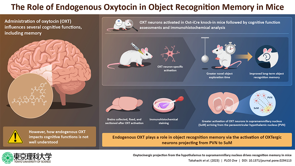
Researchers have discovered particular oxytocin neurons in the mouse brain that influence memory for object recognition.
 Oxytocinergic projection from the hypothalamus to supramammillary nucleus drives recognition memory in mice. Image Credit: Takahashi et al. (2023) PLOS One.
The hormone oxytocin (OXT) is well-known for its impact on animals' emotional bonding and psychological health. It's interesting to note that studies have revealed the critical role this brain’s natural chemical plays in learning and memory, among other cognitive functions.
By examining “OXT neurons,” which have OXT receptors and behave differently depending on the chemical's availability in the brain, scientists may have finally figured out exactly how OXT affects memory in animals.
Researchers led by Professor Akiyoshi Saitoh and Junpei Takahashi from the Tokyo University of Science examined the intricate neural pathways and signaling mechanisms triggered by OXT in a recent study that was published on November 16th, 2023, in PLOS One. They provided hitherto unseen insights into how it affects memory and learning.
Previously we had suggested that oxytocin may be a new therapeutic candidate for dementia based on studies using a mouse model of Alzheimer's disease. To investigate this further, in this study, we examined the role of endogenous OXT in mouse cognitive function. This was done by using pharmacogenetic techniques to specifically activate OXT neurons in specific brain regions. The cognitive function of mice was then evaluated using the Novel Object Recognition Task (NORT).”
Akiyoshi Saitoh, Professor, Tokyo University of Science
Because abnormal social memory in mice has been linked to deficiencies in either OXT or its receptors, the research highlights the critical role that OXT plays in regulating social memory. However, the emphasis of this ground-breaking work is now on the function of endogenous OXTergic projections in learning and memory, specifically in the supramammillary nucleus (SuM).
After specifically activating OXT neurons in the paraventricular hypothalamic nucleus (PVN), the researchers visualized mouse brain slices and observed positive signals in the PVN and its projections to the SuM.
This allowed them to identify the neurons that are responsible for OXT's effect on memory. Increased c-Fos positive cells in the PVN following the administration of clozapine N-oxide (used to activate the neurons) provided additional validation of OXTergic neuron activation.
Moreover, the Y-maze and NORT were used in the study to examine how OXTergic neuron activation affects learning and memory. Remarkably, the Y-maze test revealed no alterations in short-term spatial memory. Nonetheless, the NORT's long-term object recognition memory was greatly enhanced by the activation of OXTergic neurons.
Interestingly, after NORT, there were more c-Fos positive neurons in SuM and the dentate gyrus (a part of the hippocampus) in the brain, suggesting that OXTergic neurons are involved in preserving long-term memory through these regions.
The researchers also used selective activation of OXTergic axons in SuM, which made the mice spend more time investigating new objects. This suggests that OXTergic axons projecting from PVN to SuM directly modulate object recognition memory.
This work shows that OXT is involved in object recognition memory through the SuM for the first time. It emphasizes how OXTergic projections affect recognition memory and raises possible questions about how physiological OXT functions in Alzheimer’s disease.
There is a widely acknowledged belief that dementia tends to advance more rapidly in settings where individuals experience loneliness or limited social engagement. However, the scientific underpinnings of this phenomenon have remained largely elusive. Our research seeks to elucidate the crucial role of a stimulating environment that activates oxytocin in the brain, potentially mitigating the progression of dementia.”
Akiyoshi Saitoh, Professor, Tokyo University of Science
The continuous investigation in this domain is expected to lay the groundwork for pioneering therapies and medicinal approaches designed to impede the progression of dementia.
Source:
Journal reference:
Takahashi, J., et.al (2024) Oxytocinergic projection from the hypothalamus to supramammillary nucleus drives recognition memory in mice. PLOS One. doi.org/10.1371/journal.pone.0294113