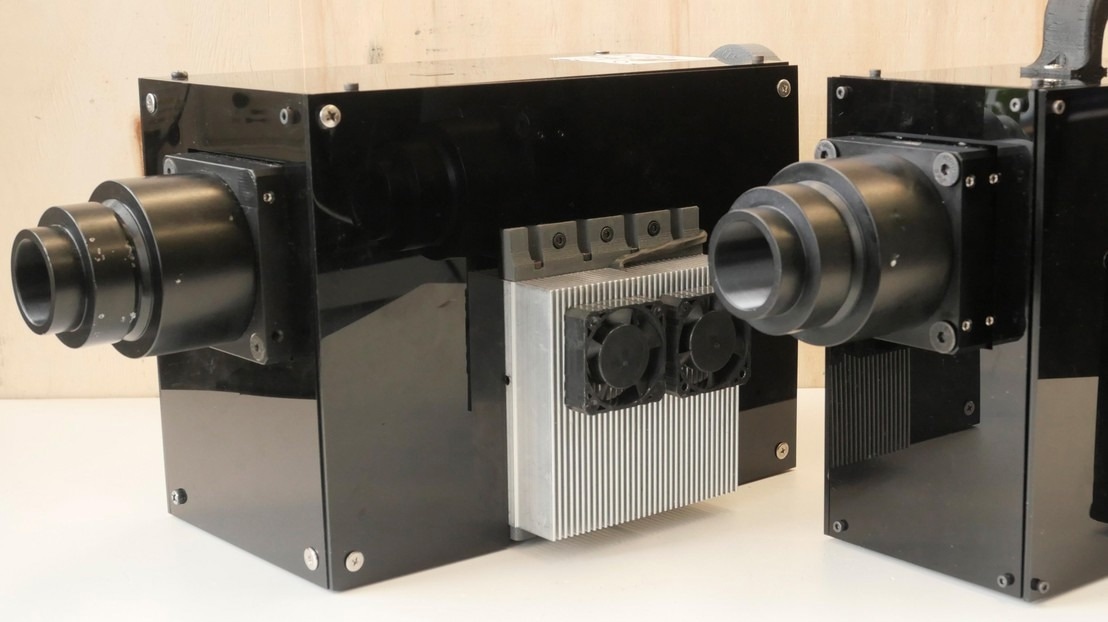
EPFL scientists have recently released a comprehensive manual detailing the construction of an add-on that can transform a regular optical microscope into a cutting-edge tool capable of generating high-resolution, three-dimensional images of cells, organoids, and embryos.

Image Credit: 2024 LBNI
For centuries, scientists relied solely on the optical microscope to investigate the motion of cells, bacteria, and yeast. However, due to the diffraction of light, it was challenging to observe objects at resolutions below 100 nm as the images produced were too indistinct to provide meaningful information.
This obstacle, referred to as the diffraction barrier, was successfully surpassed approximately 15 years ago with the introduction of super-resolution microscopy. This breakthrough enabled scientists to delve into the inner workings of living specimens, analyze organelle behavior, and witness the interactions between cells and viruses, proteins, and drug molecules.
Structured illumination microscopy (SIM) is a highly valued technique among researchers due to its ability to generate high-resolution and high-contrast images while minimizing photon exposure. Despite the advancements in electron microscopes with nanometer resolution, optical imaging remains crucial in life-science research.
It provides greater flexibility in terms of equipment and enables scientists to observe live samples under normal developmental conditions. However, the cost and limited availability of SIM imaging have hindered its widespread use. To address this issue, scientists at EPFL’s Laboratory for Bio- and Nano-Instrumentation (LBNI) within the Interfaculty Institute for Bioengineering (IBI) at EPFL’s School of Engineering (STI) have devised a method to convert a standard optical microscope into a high-resolution device using affordable, commercially accessible components. The team has published a comprehensive guide detailing the step-by-step process in open-access format, along with a series of video tutorials.
A Compact Microscope that Non-Experts Can Build and Use
The diffraction barrier is effectively surpassed by SIM through the reconstruction of the blurred regions that typically exhibit high spatial frequencies when observed using a traditional optical microscope. This technique provides a resolution enhancement of two times, allowing scientists to examine minute details measuring as small as 100 nm. SIM operates by projecting a standard illumination pattern, like a grid, onto a sample. Subsequently, images taken with various illumination patterns are processed using an algorithm to generate a higher-resolution reconstruction, utilizing the moiré effect.
In 2019, Mélanie Hannebelle, a Ph.D. student, required a microscope with specific capabilities for her research. This necessity led her to conceive the concept of constructing one for the LBNI. While other laboratories had already developed comparable devices, they were intricate, unwieldy, and challenging to replicate. Hannebelle aimed to create a more streamlined option that could be assembled and operated by individuals without expertise, without the need for costly maintenance.
We sourced electronic components of the kind used to make the video projectors you see in classrooms. We altered and arranged them, so they were capable of projecting a light pattern onto a sample.”
Georg Fantner, Professor, LBNI
Tested and Approved By Life-Sciences Researchers
The LBNI team sought to determine if their new microscope could serve as a feasible and practical option. Consequently, they enlisted the help of various laboratories to conduct tests. Collaborating with Professors Andrew Oates, Matthias Lutolf, John McKinney, and Aleksandra Radenovic, they evaluated the instrument using authentic research samples.
Our colleagues asked us questions, told us about their needs, and shared their samples with us. We were eager to find out whether and how our instrument could help them in their research.”
Georg Fantner, Professor, LBNI
The response to the project was predominantly positive, leading the team to obtain an EPFL Open Science grant to disseminate their instrument in an open-hardware format. Transforming the device into a reproducible tool for other laboratories required a meticulous and time-consuming effort.
Esther Raeth, a fellow Ph.D. student in the same laboratory, compiled comprehensive instructions, equipment lists, and video tutorials to be published online.
Fatner added, “The only prerequisite for our system is a high-quality optical microscope – something most labs already have.”
The OpenSIM is not designed to rival more advanced tools. While it may have a lower modulation contrast compared to commercially available options, limiting the resolution-gain to 1.7× instead of the theoretical 2x, it still fulfills its intended role. This allows labs with occasional needs or budget constraints to access SIM technology without having to invest in a high-end model costing CHF 500,000 or more. The LBNI team is committed to expanding the reach of their work to a broader audience of scientists and creating a community of users to exchange knowledge and insights.
Prof. Fantner concluded, “Since the paper was shared on BioRxiv.org, I’ve been contacted by several people who are interested in the idea and want to know more about how to build their own OpenSIM.”
Source:
Journal reference:
Hannebelle, M.T.M., et al., (2024) Open-source microscope add-on for structured illumination microscopy. Nature Communications. doi.org/10.1038/s41467-024-45567-7