Modern high-content screening microscopes enable fast, automated imaging of whole microtiter plates by imaging fixed positions inside different wells. This is perfect for in-vitro cell culture-based readouts and other tests with evenly distributed phenotypes. However, it becomes limited when large specimens or rare events are examined, as the limited field of view (FOV) of high-magnification objectives might not fully encompass the area of interest.
Researchers are frequently limited to lesser magnification acquisition, resulting in data with low resolution or the omission of features of interest in multiple wells. This article explains how Bruker's Acquifer Imaging Machine (IM) overcomes these issues with PlateViewer software and provides a semi-automated technique for experiments involving advanced supervised feedback microscopy.
Automated Experimental Approaches
For tissue-specific imaging in large specimens (e.g., zebrafish), a method that can automatically identify and zoom in on the tissue or organ of interest is required. Fully automated tissue detection and imaging frequently require complex image processing routines to be developed. Each project requires careful balancing between software development time and project size.
Semi-automated methods represent an optimal solution, requiring minimal user interaction and no custom algorithm development. This simpler alternative can easily handle even complicated or variable structures that would typically demand extensive development of image detection routines. This method is useful for screening projects as they start instantly, saving both time and resources.
Plate-Viewer Software

Figure 1. Illustration of Click-Tool functionality. (a) Zebrafish embryo imaged with a 2x objective and visualized in Plate-Viewer. The crosshair is centered on the heart region in a three-day-old embryo of the epi:GFP;myl7mR transgenic line. The red bounding box indicates the FOV of a 10x objective used for subsequent high-resolution imaging. (b) Single Z-plane of a high-resolution dataset is automatically acquired on Acquifer IM. Image Credit: Bruker Nano Surfaces and Metrology
Plate-Viewer software provides a semi-automated method for supervised feedback microscopy. Low-magnification pre-screen data of a full microtiter plate is visualized after the plate layout for a rapid and easy-to-understand overview. The integrated “click-tool” function permits assay experts to select areas of interest (ROIs) for different wells.
Furthermore, in-built and generic “template-matching” algorithms facilitate robust automatic localization of several target structures. These include complicated reporter expression patterns, morphological features, and rare events in different wells. Acquifer IM spontaneously gathers information at a high resolution from specific areas that correspond to predefined settings, and acquired high-resolution data is then easily and intuitively visualized in Plate-Viewer.
Plate-Viewer has multiple distinct properties that make it possible for users to:
- Select ROIs to be automatically imaged by the click of a mouse or template matching
- Preview FOVs of higher magnification objectives via an adjustable bounding box
- Intuitively visualize and search through large-scale screening datasets
- Easily search through sophisticated multidimensional datasets
- Apply lookup tables and overlay channels to enhance manual inspection
- Change basic image parameters to improve the visualization of details
- Plug-in interface for integration of external image processing instruments
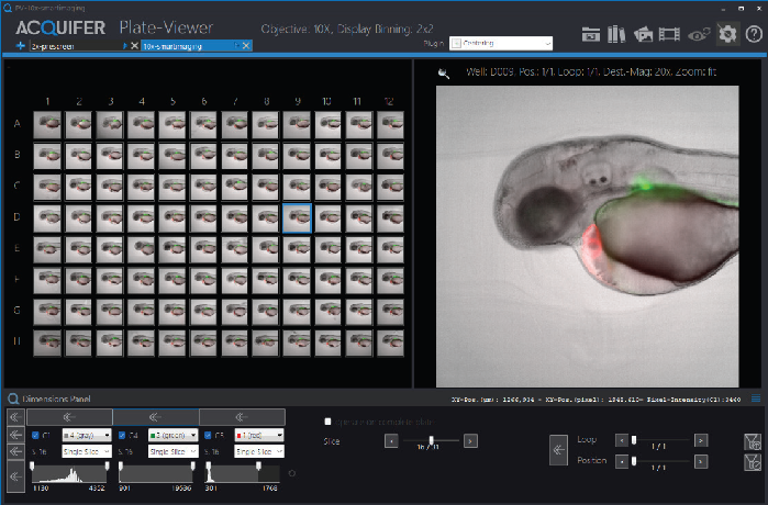
Figure 2. Overview of the Plate-Viewer functionalities. Visualization of identical planes of all available wells within a microtiter plate (left). Preview of the currently selected well coordinate (right). Control panels in the top-right corner enable the generation of colored overlay images and the display of selected channels. Control panels in the lower left corner allow channel-specific adjustment of the histogram and the choice of navigation mode through multidimensional datasets (e.g., slice, loops, or subpositions). Image Credit: Bruker Nano Surfaces and Metrology
Intuitive Solution for High-Content Screening
Acquifer IM is a complete solution for experiments involving supervised feedback microscopy. Its Plate-Viewer software makes use of a semi-automated method to simplify complicated high-content screening experiments. This dynamic tool allows researchers to more easily and reliably carry out tissue-specific imaging of complex and variable structures in large specimens.
About Bruker Nano Surfaces and Metrology
Bruker Nano Surfaces and Metrology provides high-performance, specialized analysis, and testing technology for the widest range of research and production applications.
Our broad portfolio of 2D and 3D surface profiler solutions supply the specific information needed to answer R&D, QA/QC, and surface measurement questions with speed, accuracy, and ease. And our tribometers and mechanical testers deliver practical data used to help improve development of materials and tribological systems. Bruker’s industry-leading quantitative nanomechanical and nanotribological test instruments are specifically designed to enable new frontiers in nanoscale materials characterization, materials development, and process monitoring.
Bruker has been leading the expansion of atomic force microscope (AFM) capabilities since the very beginning, and our systems are the most cited AFMs in the world. Our comprehensive suite of AFMs enable scientists around the world to make discoveries and advance their understanding of materials and biological systems. With our nanoIR technology, Bruker is now also the recognized leader in photothermal IR spectroscopy from the nanoscale to the sub-micron and macro scales. And, as the only AFM manufacturer with a state-of-the-art probes nanofabrication facility and world-wide, application-specific customer support, Bruker is uniquely positioned to provide the equipment, guidance, and support for all your nanoscale research needs.
Bruker’s suite of fluorescence microscopy systems provides a full range of solutions for life science researchers. Our multiphoton imaging systems provide the imaging depth, speed and resolution required for intravital imaging applications, and our confocal systems enable cell biologists to study function and structure using live-cell imaging at speeds and durations previously not possible. Bruker’s super-resolution microscopes are setting new standards with quantitative single molecule localization that allows for the direct investigation of the molecular positions and distribution of proteins within the cellular environment. And our Luxendo light-sheet microscopes, are revolutionizing long-term studies in developmental biology and investigation of dynamic processes in cell culture and small animal models.
In addition to developing and manufacturing next-generation systems to help our customers’ current and future applications, Bruker is also very active in acquiring and partnering with innovative companies to continue to expand our range of enabling technologies and solutions. Recent additions to the Bruker Nano Surfaces family include Alicona Imaging, Anasys Instruments, Hysitron, JPK Instruments, and Luxendo.
Whatever your measurement and analysis needs, whatever your material or scale of investigation, Bruker has a specialized high-performance solution for you.
Sponsored Content Policy: AZoLifeSciences publishes articles and related content that may be derived from sources where we have existing commercial relationships, provided such content adds value to the core editorial ethos of AZoLifeSciences which is to educate and inform site visitors interested in life science news and information.