Flow cytometry is an invaluable tool used to analyze the chemical and physical properties of cells. This laboratory technique uses an antibody conjugated with a fluorochrome for cell analysis.

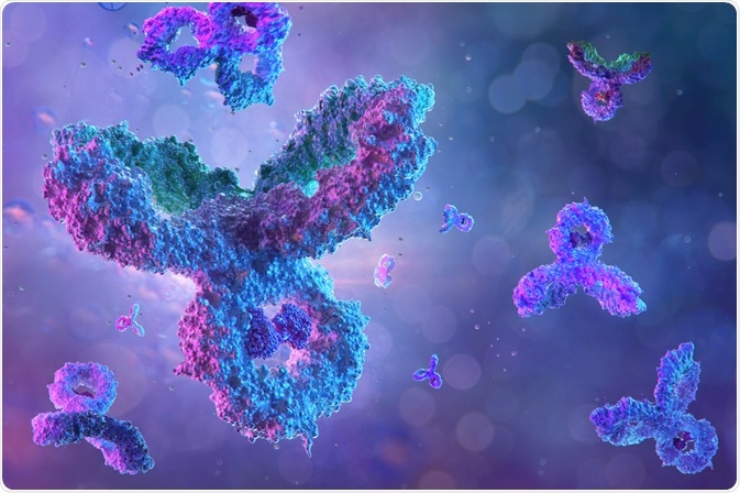
Antibodies. Image Credit: Corona Borealis Studio/Shutterstock.com
The development of a specialized flow cytometer in the 1970s, triggered by the HIV pandemic, paved the way to a broad range of applications such as protein modification studies, immunophenotyping, and analysis of intracellular antigens. The most important clinical application of flow cytometry is in hematologic malignancy diagnosis.
Principles of flow cytometry
In flow cytometric analysis, suspension of a single cell or particle is prepared and aspirated into a flow cell. The particles are made to pass through a focused laser beam one at a time and the light is either absorbed or scattered by the cells. Antibodies labeled with fluorochromes are attached to the cell surface, which helps the cells re-emit absorbed light as fluorescence.
The fluorescence signals are received by an array of photodiodes and amplified. The electrical pulses that are formed are converted into digital data that can be analyzed, displayed, and stored in a computer. Thus, statistically valid quantitative data related to a huge number of cells can be obtained in a very short period of time.
Role of antibodies in flow cytometry
Antibodies are an invaluable component of flow cytometry. The advent of monoclonal antibodies in 1977 promised an unlimited supply of highly specific antibodies and dramatically changed the flow cytometry technique. Monoclonal antibodies are produced from single B-cell clones developed in hybridoma cells.
They have homogeneous antigen-binding sites and therefore, are highly specific to antigenic determinants. Over the years, specific monoclonal antibodies for murine MHC antigens and murine/rat helper T cells have been developed. The identification of 3 murine monoclonal antibodies that are specific for human T-cells - OKTl, OKT3, and OKT-l-lI7 – opened up new avenues for human studies.
Monoclonal antibodies generated against a wide range of biological molecules such as glycoproteins, proteins, carbohydrates, glycolipids, histones, lysosomes, and cytokines have been produced over the years.
The development of monoclonal antibodies against specific phosphoepitopes that can help in the detection of protein activation states has enabled the use of flow cytometry to study cellular function. Since antibodies can recognize and bind to antigenic epitopes, they can be used to identify different molecules with specific epitopes.
The size and shape of the antibody used and its conjugates influence the staining measurements in flow cytometry, especially in the case of cytoplasmic or intranuclear staining. Controlled permeabilization is crucial to facilitate the penetration of the fluorochrome-antibody conjugate into the nucleus or the cytoplasm and to achieve optimum extracellular staining. Also, high concentrations of antibodies in flow cytometry can lead to non-specific binding which will complicate the process.
Antibody stability is another concern that affects staining in flow cytometry. Exposure to extreme salt concentration or pH decreases the stability of antibodies and they tend to form soluble aggregates and precipitated polymers.
This is probably because of their increased hydrophobicity, which increases the possibility of non-specific binding. Therefore, these aggregates and polymers need to be removed from antibodies before using them in flow cytometry. All these factors need to be carefully considered before the formulation of antibody-conjugates for flow cytometry.
Sigma® Life Science, the Life Science division of Sigma-Aldrich, provides monoclonal antibodies validated specifically for flow cytometry in pure form or conjugated to some commonly used fluorochromes. The company offers antibodies against MHC and related antigens, immunoglobulins, and CD and related antigens.
Thermo Fisher Scientific offers antibodies conjugated to 24 different fluorescent dyes and proteins for use in flow cytometry. This allows more flexibility in experiment design and expands the range of the detectable parameter of flow cytometry.
References
Further Reading
Last Updated: Jul 1, 2022