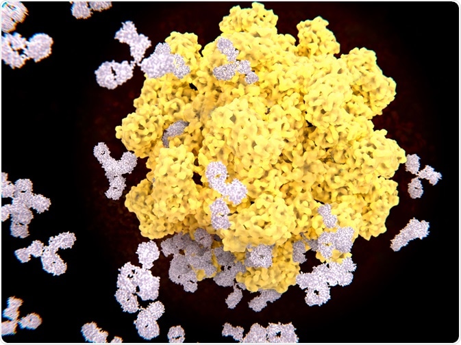
There are two types of immunity: innate and adaptive immunity. Innate immunity is developed in utero and present at birth. Adaptive or acquired immunity is developed as an individual is exposed to antigens.

Image Credit: Juan Gaertner/Shutterstock.com
The innate immune system is the primary defense mechanism against pathogens. It is comprised of non-specific defense mechanisms such as; the skin and hair (physical barrier), secretions (mucous, gastric acid, sweat), and general immune responses (non-specific cell responses, inflammation, complement). Part of the innate immune system's role is to activate the acquired immune system.
The acquired immunity is highly specific to a particular antigen via the production of antibodies, creating an immunological memory after an initial response to a specific pathogen. The main function of an antibody is to bind specifically to their target antigen, eliciting an immune response against the bound antigen by recruiting other cells and molecules.
The interaction between antibody and antigen involves numerous weak noncovalent bonds between the paratopes (the binding site on the antibody) and epitope (the binding site on the antigen).
Antigen processing and presentation
Antigen processing, also known as the cytosolic pathway is part of the immunological pathway which prepares antigens of non-self cells/pathogens to the specialized cells, T-lymphocytes. There are two pathways in which the immune system differentiates the antigens from self and presents them through class I or II major histocompatibility complex (MHC).
It is a prerequisite that MHC I and MHC II both bind to the antigen before it can be presented on the cell surface membrane. These two pathways are known as endogenous and exogenous pathways.
The endogenous pathway presents the fragments of the cell proteins on the cell surface via MHC I. This flags the cell for destruction, allowing the immune system to recognize the cell as infected. The exogenous pathway is the pathway which antigen-presenting cells use to present the antigens obtained from endocytosed proteins, this functions through MHC II.
Antigen-presenting cells are a heterogeneous group of immune cells, comprising of dendritic cells, macrophages, and B cells. These immune cells initiate the cellular immune response by engulfing pathogens and processing antigens, presenting the processed antigens in conjunction with MHC class II molecules on the cell surface where it can interact with helper T cell receptors. This leads to the activation of helper T cells. The processed antigen part presented by MHC is called an epitope.
The activated helper T cells have a role in the activation of cytotoxic T cells to kill infected target cells, macrophages to attack infected cells, and B cells to proliferate and differentiate into either memory cells or plasma cells (antibody-secreting effector cells).
When naïve or memory B cells are activated by antigens (with the aid of helper T cell), they proliferate and differentiate into plasma cells. Such cells produce a great amount of soluble (not membrane-bound) antibody, which has the same unique antigen-binding site as the cell-surface antibody, working earlier as the antigen receptor.
Although many plasma cells die after several days, some survive in the bone marrow for months or years, still possessing the ability to secrete antibodies into the blood when necessary.
Antibody binding site
Antibodies are composed of four polypeptide chains: two identical heavy (H) chains and two identical light (L) chains. The light and heavy chains are connected by disulfide bonds, forming a similar shape to the letter "Y" with two arms and a single base.
Each arm is known as the Fab domain, and the single base is known as the fragment crystallization region or Fc domain. Thus, a single antibody consists of two Fab domains and one Fc domain.
The Fab consists of two variable domains (VH in the heavy chain and VL in the light chain) and two constant domains (CH1 and CL), with the two variable domains (VH and VL) making up the variable fragment (Fv), which provides the antigen specificity of the antibody. The constant domains of Fab act as a structural framework.
Six hypervariable loops lie within each variable fragment: three in the light chain (L1, L2, and L3) and three in the heavy chain (H1, H2, and H3), supported by a conserved framework region of β-sheets. The variable domain consists of seven amino acid regions: three hypervariable regions and four conserved framework regions.
The six hypervariable loops within each variable fragment, commonly known as complementarity determining regions (CDRs), are widely considered to be responsible for antigen recognition.
The heavy and light variable domains fold in a specific way that brings the hypervariable loops together to form the antigen-binding site or paratope. While the framework regions show high degrees of similarity, the CDR regions are characterized by their divergence.
Antibodies have a unique ability to bind virtually to any “non-self” surface with superior specificity and high affinity, making them not only the key to immunity but also an extremely valuable tool in experimental biology, biomedical research, diagnostics, and therapy.
Sources
- Blum, J.S., Wearsch, P.A., and Cresswell, P., 2013. Pathways of antigen processing. Annual review of immunology, 31, pp.443-473.
- Brodsky, F.M., and Guagliardi, L.E., 1991. The cell biology of antigen processing and presentation. Annual review of immunology, 9(1), pp.707-744.
- Davies, D.R., and Chacko, S., 1993. Antibody structure. Accounts of chemical research, 26(8), pp.421-427.
- Edelman, G.M., 1973. Antibody structure and molecular immunology. Science, 180(4088), pp.830-840.
- Germain, R.N., and Margulies, D.H., 1993. The biochemistry and cell biology of antigen processing and presentation. Annual review of immunology, 11(1), pp.403-450.
- Janeway, C.A., Travers, P., Walprot, M., and Shlomchik, M., 2001. Part II: recognition of antigen. Immunobiology, 5th ed., Garland Science, New York, and London.
- Sela-Culang, I., Kunik, V., & Ofran, Y. (2013). The structural basis of antibody-antigen recognition. Frontiers in immunology, 4, 302.
- Van Regenmortel, M. H. (2019). Specificity, polyspecificity, and heterospecificity of antibody-antigen recognition. In HIV/AIDS: Immunochemistry, Reductionism, and Vaccine Design (pp. 39-56). Springer, Cham.
Further Reading