Biomolecule imaging has been revolutionized in recent years by advancements in cutting-edge cryogenic electron microscopy (cryo-EM) technology. As a result, scientists have been able to visualize the structure of small biomolecules such as viruses and proteins in unprecedented levels of detail. This has opened up numerous avenues to explore in terms of the development of new therapeutic options for various diseases.

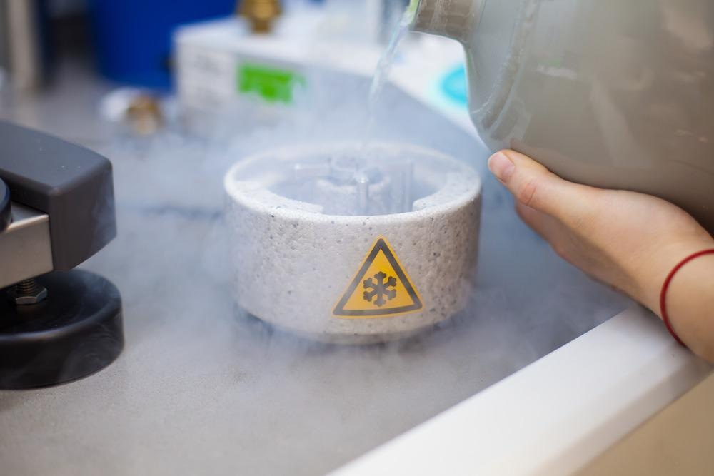
Image Credit: PolakPhoto/Shutterstock.com
However, the field had been prevented from reaching its full potential due to the high cost to the technology, the expertise required to operate it, and the relatively lower resolution it offers in comparison to alternative methods such as X-ray crystallography. These factors had forced cryo-EM to remain inaccessible to many research teams until recent developments established cryo-EM technology that is low-cost and easy-to-use, as well as microscopes that can produce images with atomic resolution.
Here, we provide an overview of the technology, detail the challenges it faces, and explain how recent development are overcoming these challenges. Finally, we predict how cryo-EM will develop into the future.
What is cryogenic electron microscopy?
Cryo-EM is an imaging technique used to produce high-resolution three-dimensional reconstructions of biomolecules. It works by flash-freezing biological samples in glass-like ice and using a high-energy electron beam to probe the specimen. Upon firing the electrons at the sample, the atoms within the sample scatter and change their direction. These scattered electrons are then picked up by detectors, allowing the unique scatter pattern to be mapped to create an image of the biomolecule sample.
While the method provides high-resolution three-dimensional reconstructions, for many years, the images it has produced have fallen second to those produced by X-ray crystallography, which is able to produce much higher-resolution images. While the method of cryo-EM is easier than that of X-ray crystallography in some ways, X-ray crystallography has generally been favored due to its ability to better visualize biological structures.
Additionally, for many years cryo-EM has been difficult to implement in many laboratories due to the high cost of the equipment and the need for highly trained staff to operate it. As a result, the technology has failed to become widely adopted. Fortunately, this is all set to change with the recent advances that have been achieved in the field.
Overcoming challenges in cryogenic electron microscopy
A team of researchers at the Biodesign Center for Applied Structural Discovery (CASD) and ASU's School of Molecular Sciences (SMS) have refined the technique of cryo-EM, enabling it to produce higher resolution images.
Proteins, such as those that are often the target of cryo-EM investigations, have unique three-dimensional structures that determine their functioning. It is possible to take snapshots of these structures in different conformations, some of which may persist in time, while others are ephemeral, existing for just billionths of a second, making them challenging to capture.
The new research conducted at CASD and ASU has established an updated method of cryo-EM that models these transitory structures by capturing and including images of these fleeting conformations rather than producing the ‘average’ image which only captures the configurations that endure over time. This advance in technology helps overcome the resolution limitation of cryo-EM.
In another recent study conducted at the Okinawa Institute of Science and Technology (OIST) Graduate University, scientists have established a more cost-effective and easier to operate cryo-electron microscope.
In traditional cryo-EM, electrons from an electron beam interact with the atoms of a biological specimen, creating scatter which is picked up on by detectors to build an image of the sample’s structure. Using high-energy electrons causes just a small number of atoms to scatter due to the weak interactions between the atoms of low atomic mass (such as carbon, nitrogen, hydrogen, and oxygen) and the electrons. Low energy electrons, on the other hand, are slower and interact more strongly with the atoms, producing more frequent scattering. Using low-energy electrons is more cost-effective, but until recently, this method had been inappropriate due to the significant amount of background noise it generated via the scattering caused by the surrounding ice of the specimen.
Scientists at the OIST have overcome this limitation by adapting a cryo-EM microscope that can switch to cryo-electron holography. The process of cryo-electron holography is quick and simple, overcoming the previous challenges of cryo-EM.
The future of cryogenic electron microscopy
Given that the biological specimens imaged with cryo-EM, such as proteins, are often the primary targets for pharmaceutical drugs, developments in this field are vital to furthering our understanding of disease. It is likely that in the coming years, more research and development will continue to improve the technique of cryo-EM, helping to facilitate its widespread adoption and further its capabilities. The technique will likely be involved in future discoveries that will be key to the development of new therapeutics for several diseases.
Sources:
- Callaway, E., 2020. ‘It opens up a whole new universe’: Revolutionary microscopy technique sees individual atoms for first time. Nature, 582(7811), pp.156-157. https://www.nature.com/articles/d41586-020-01658-1
- Cheung, M., Adaniya, H., Cassidy, C., Yamashita, M. and Shintake, T., 2020. Low-energy in-line electron holographic imaging of vitreous ice-embedded small biomolecules using a modified scanning electron microscope. Ultramicroscopy, 209, p.112883. www.sciencedirect.com/.../S030439911930289X?via%3Dihub
- Shekhar, M., Terashi, G., Gupta, C., Sarkar, D., Debussche, G., Sisco, N., Nguyen, J., Mondal, A., Vant, J., Fromme, P., Van Horn, W., Tajkhorshid, E., Kihara, D., Dill, K., Perez, A. and Singharoy, A., 2021. CryoFold: Determining protein structures and data-guided ensembles from cryo-EM density maps. Matter, 4(10), pp.3195-3216. www.cell.com/.../S2590-2385(21)00449-5
Further Reading