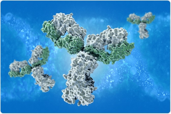
Paratopes and epitopes are the unique binding regions of an antibody and antigen, respectively. More specifically, antigens are known to contain specific antigenic determinants (which are epitopes), while antibodies contain antigen-binding sites (which are paratopes).

Image Credit: vitstudio/Shutterstock.com
What is an epitope?
The binding of an antibody to a foreign body occurs across a specific region that is a few amino acids long and does not represent the whole protein. This region, which is recognized by the antibody, is called the antigen. A protein may often contain several epitopes to which antibodies may bind.
There are two types of epitopes: continuous and discontinuous. Continuous epitopes are a linear sequence of amino acids that are recognized by the antibody. Discontinuous epitopes are regions that are present only in the folded conformation of a protein.
The type of epitope present determines the antibody type. Monoclonal antibodies are only able to recognize one epitope, whereas polyclonal antibodies recognize multiple epitopes.
Epitope mapping
There are two methods that have been widely used to study the structure of epitopes: x-ray crystallography and monoclonal antibodies.
Since polyclonal antibodies recognize different epitopes and with different affinities, it is difficult to identify the map the location of epitope using polyclonal antibodies. In addition, as different polyclonal antibodies may recognize overlapping epitopes, it is hard to correspond a specific polyclonal with a specific sequence.
Methods such as SDS-PAGE cannot be used as this method partially denatures the protein, which can modify the antibody-epitope recognition. Techniques such as liquid phase immunoassays and frozen tissue samples often simulate the in vivo affinities. Different monoclonal bodies can be used to pinpoint different regions of the epitope; furthermore, disulfide bridges can also be identified by mapping the epitope.
X-ray crystallography is a highly precise method to map the tertiary structure between the epitope and the corresponding antibody. However, this method is expensive and requires the sample to be in crystal form.
Nuclear magnetic resonance (NMR) can also used for this purpose; however, it has somewhat lower precision than X-ray crystallography, and the size of the antigen is a limiting factor. For larger sized antigens, electron microscopy can also be used.
What is a paratope?
A paratope is the region of antibody that recognizes and binds to the epitope of an antigen. Paratope are produced by the complementary binding of light and heavy chains that create a three-dimensional structure. In theory, 104 heavy chains can combine with 104 light chains to generate 108 different paratopes.
The paratope region is present in the Fv region of the antibody and consists of 5-10 amino acids. To determine the precise structure of paratope, the structure of the antibody complexed with the antigen should be determined, and this can be done using co-crystallography and structural elucidation.
The epitope region to which the paratope binds can often be mimicked by macromolecules that are similar to epitope - such molecules are termed mimotope.
Paratope structure prediction
Currently, software that use antibody statistics to assign a score to different residues are presently based on how likely they are to be in contact with an antigen. High score indicates high probability of the residue to be a part of the paratope sequence, while a low score indicates a low probability of being a part of paratope. Such scores help in predicting the paratope regions of an antibody.
Further Reading
Last Updated: Sep 8, 2022