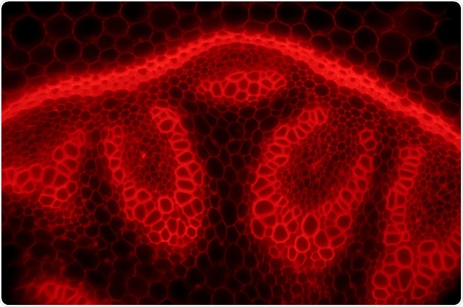
Epifluorescence microscopes are a commonly used tool for studying specimens. Improved specificity and contrast provided by the application of fluorescence to the field of microscopy has stimulated the advancement of various discoveries in the biosciences.

Image Credit: D. Kucharski K. Kucharska/Shutterstock.com
In 1843, George Gabriel Stokes described fluorescence as being characterized by the wavelength of an emitted light that is longer than the wavelength of the exciting light.
This discovery led to the invention of the epifluorescence microscope, now a ubiquitous instrument in biological and medical laboratories.
The high contrast and specificity of the images produced have allowed for the increased understanding of cell structures, the location, and dynamics of gene expression and distinct interactions at a molecular level within the cell.
Principles of fluorescence
Fluorescence microscopy is a particular form of light microscopy in which, instead of utilizing visible light to illuminate specimens, a higher intensity light source excites a fluorescent molecule called a fluorophore.
The fluorophore absorbs photons leading to electrons moving to a higher energy state. When the electrons return to the ground state by losing energy, the fluorophore emits light of a longer wavelength.
The emitted light is then separated from the original excitation light via a filter and this produces a magnified image of the specimen being studied.
The use of fluorescence allows for the object of interest to be specifically targeted. As the excitation light is filtered out, the emitted fluorescence allows only the fluorescent objects of interest to remain visible.
Components of an epifluorescence microscope
An epifluorescence microscope requires particular components to separate the excitation and emission lights, acquire the image and allow for the target object to be observed. The following are important components of an epifluorescence microscope:
- The light source: A source of high-intensity light that can emit a broad spectrum of wavelengths is required. This is usually in the form of a xenon arc lamp or a high-pressure short arc mercury lamp.
- Filter cubes: They allow for the selection of specific excitation and emission wavelengths and consist of the excitation filter, dichroic mirror, and barrier filter. The transmission of the excitation light is selected via the excitation filter and the transmission of the emission light is selected via the barrier filter, by blocking the excitation light. The dichroic mirror separates the excitation light from the emission light further by reflecting the shorter wavelengths of the excitation light and transmitting the longer wavelengths of the emission light.
- Objective lens: The objective lens transmits light to the sample to form the image. The emission light passes down through the dichroic mirror before reaching the objective lens.
- Camera system: Modern epifluorescence microscopes can record the images of the specimen with high-resolution. Electron multiplying CCD cameras are often used as they are capable of imaging single-photon events without requiring an image intensifier. This means that images can be captured at high speed without loss of sensitivity.
Epifluorescence illumination or epi-illumination
Epifluorescence illumination or epi-illumination is the specific arrangement of these optical components that allow for the illumination of the specimen from above. The objective lens acts as both the source of light that excites the fluorophore and the element of the microscope that collects the emitted fluorescent light.
In comparison to other forms of fluorescence microscopy, epifluorescence illumination has the advantage of only requiring a small amount of emitted light to be blocked.
Though this approach means that the excitation light and emission light overlap, the dichroic mirror efficiently separates the excitation from the emission.
The combination of an excitation filter, dichroic mirror, and barrier filter in the filter cubes means that only one photon in 10,000 of the wrong excitation wavelength to the specimen passes, with a similar amount for the emission light. This high ratio is important for clearly imaging single fluorescent molecules.
Terminology:
- Fluorophore: A chemical compound that fluoresces by emitting light upon excitation.
- Photon: A fundamental particle of visible light.
- Ground state: The normal, non-excited state of a molecule.
- Excited state: Electrons can move to a higher energy level by absorbing light, therefore achieving an excited state.
Sources:
- Masters, B.R. 2010. ‘The Development of Fluorescence Microscopy’. In: Encyclopedia of Life Sciences (ELS). John Wiley & Sons. http://onlinelibrary.wiley.com/doi/10.1002/9780470015902.a0022093/abstract
- Webb, D. and Brown, C. 2013. ‘Epi-Fluorescence Microscopy’, Methods in Molecular Biology, 931, pp. 29-59. https://www.ncbi.nlm.nih.gov/pmc/articles/PMC3483677/
- www.ammrf.org.au/.../index.php
- Lichtman, J. and Conchello, J. 2005. ‘Fluoresence microscopy’, Nature Methods, 2, pp. 910-913. https://microscopy.berkeley.edu/
- https://microscopy.berkeley.edu/
Further Reading