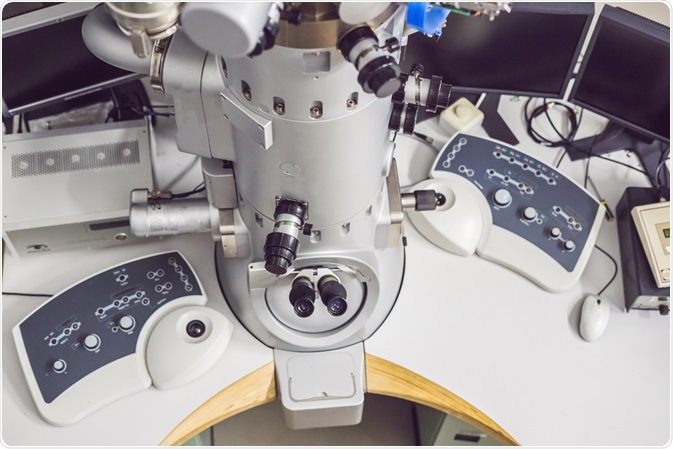
Since the invention of the electron microscope in 1931, scientists have been able to investigate the hidden world of the nanomolecular. Over the years, the electron microscope has been improved upon, with a wide array of transmission electron microscopy (TEM) imaging modes now available for researchers to use.
 Image Credit: Elizaveta Galitckaia / Shutterstock.com
These include conventional imaging, STEM (scanning transmission electron microscopy) imaging, spectroscopy, diffraction, and combinations of these imaging modes. In conventional imaging alone, contrast can be produced in many different fundamental ways, referred to as “image contrast mechanisms.”
Different modes of imaging can be achieved by selecting different parts of the electron microscope, such as the objective aperture or lens, which give differing results. Some imaging modes are listed below.
Bright-field imaging
Bright-field imaging is the most commonly used conventional imaging mode. In this technique, transmitted electrons are selected by the aperture to provide an image, resulting in the sample appearing light and the background appearing dark. This is a straightforward method.
Dark-field imaging
Dark-field imaging is effectively the opposite of bright-field imaging. The aperture is used to select the scattered electrons, with the transmitted electrons blocked. This provides an image where the sample in the foreground appears dark, whereas the background appears light, which allows the study of structures such as crystal lattices, which may be too small for bright-field imaging to pick out.
There are a few methods that can be employed by the electron microscope operator to produce a dark-field image. These include:
- The objective aperture is moved, which excludes the unscattered beam whilst allowing some of the signal to pass through the area of interest (for example, a particular intensity spot.)
- The operator tilts the beam, blocking the path of the unscattered beam by the objective aperture. In so doing, a specific intensity spot can be centered, becoming the new “main beam.” This means that the dark-field image is produced only from the electrons, which are diffracted along this axis. The image is then focused on the sample.
Diffraction imaging
The result of Bragg scattering caused by a beam passing through a crystalline structure, diffraction images differ from conventional TEM images.
By inserting a “selected area diffraction aperture” and delimiting the region of interest, an image is created in the back focal plane (below the sample), which is seen as either a set of diffuse rings or an array of dots. The crystal structure can be extrapolated from these images.
Convergent Beam Electron Diffraction (CBED)
In CBED, instead of passing a stream of electrons through the sample, diffraction patterns are recorded using a conical beam focused, and so it converges to a single point on the sample. Information about the specimen’s crystal symmetry can be extrapolated from the discs that are formed by the diffraction pattern. This is achieved by adjusting the focus (objective lens) rather than the objective aperture to bring the sample into focus.
Phase-contrast or high-transmission electron microscopy
Also known as high-transmission electron microscopy (HRTEM), phase contrast can be used to investigate crystal structure. Images are formed in this process due to the differences in the electron wave phase caused by specimen interaction.
The image is dependent on the number of electrons that hit the screen, so the interpretation of phase contrast images can be complex. This effect, however, can be used to advantage by the skilled operator, as it can be manipulated to give more information about the sample, such as in phase retrieval techniques.
3D-TEM
Three-dimensional transmission electron microscopy provides, as the name suggests, 3D images of a sample. This method uses multiple imaging of a sample combined with image analysis and provides structural information of thin samples with a clarity that is not available with conventional imaging modes.
TEM imaging modes: multiple possibilities
Electron microscopy is a dynamic science with many standards and non-standard imaging methods. In the hands of a trained operator, the possibilities for study and analysis of the micromolecular world are many, with various imaging modes available, and more being developed as the technology progresses. This article has been an introduction to just some of the imaging modes available in the field of transmission electron microscopy today.
Further Reading