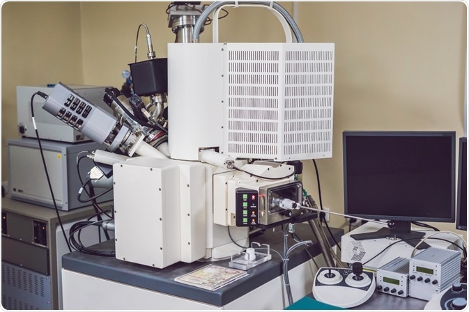
FIB-SEM can be thought of as an enhanced version of SEM that produces high-resolution images, being used in several applications across industries.
The method continues to be developed as it finds more uses, from generating high-quality 3D images, to visualizing neurons and helping develop fuel cell technology.

Image Credit:Elizaveta Galitckaia/Shutterstock.com
FIB-SEM (Focused Ion Beam Scanning Electron Microscope) is a technology developed from the simpler form of SEM (Scanning Electron Microscope).
The difference between SEM and FIB-SEM is that the beam of electrons that is used in SEM is replaced by a focused beam of ions.
The two techniques are almost identical, apart from the switch of the electron beam to a focused beam of ions. Also, the FIB-SEM technology produces images that have much higher resolutions to the nanometer-level.
In FIB, a focused beam of ions is used to directly affect the surface of a sample. Both the energy and the movement of the beam are precisely controlled with nanometer accuracy to achieve a certain effect, such as creating minute components or removing ultrathin layers of tissue/material for analysis.
A FIB becomes even more powerful when it is combined with an SEM, where a dual electron beam intersects the ion beam at a 52° just above the sample’s surface, producing instantaneous SEM imaging of the surface milled by FIB. The FIB-SEM technique combines precise, high-resolution imaging with chemical analysis capabilities.
The method has been widely adopted in the fields of semiconductor and electronic development, materials science, biology, neuroscience, and more.
Below, we discuss the major applications of FIB-SEM technology.
The applications of FIB-SEM
3D imaging
The 3D imaging capability of the FIB-SEM technique has made it an attractive investigative technique adopted for many industries to be used in a wide range of applications. The images are produced with a superior z-axis resolution, meaning that processes such as registration and post-processing are not required as much as with comparable techniques.
Recent research has helped to develop FIB-SEM so that it can overcome its drawbacks of relatively slow imaging speed and lack of stability which up until recently, had limited the possible acquisition volume of the method, therefore capping its use in different applications.
A recent study, published in the journal Materials Science in Semiconductor processing, revealed how scientists had been able to expand these volumes while improving the reliability of FIB-SEM. The research team had been able to expand the FIB-SEM method, which was shown to be particularly effective at producing higher resolutions on smaller volumes.
This step forward opened up several new applications of FIB-SEM, including the examination of neural tissue and the exploration of cell biology.
Semiconductor development
FIB-SEM has been adopted by the electronics sector to help develop better functioning semiconductors. The technology is being used to scan and characterize the 3D structures of the semiconductor devices under development, to help researchers better understand how to predict and prevent component failure and improve on their designs.
Because of FIB-SEM’s high level of accuracy, plus its fast turnaround time, it has become very popular in this application due to the high-pressure demands of the sector. Within a matter of hours, in-depth structural information can be produced.
Research continues to explore ways in which FIB-SEM can be used to help improve semiconductor devices.
Recent studies have used FIB-SEM to analyze defects and establish failure localization of materials used in superconductors. This helps scientists to create materials that are more reliable, efficient, and better performing.
Visualizing neurons
Biology has also greatly benefited from the use of the FIB-SEM technique which has been adapted for use in the analysis of biological samples. This has been particularly useful for the exploration of neurons.
As the resolution of FIB-SEM is at the level of the nanometer, scientists have been able to use this technique to not only view the structure of neurons, but also gain insight into how they work, how they respond to stimuli, and how they grow and adapt.
FIB-SEM may continue to be used in this field to elucidate key information about the nature of neurons, which may lead to the development of new treatments for a multitude of diseases and disorders, both mental and physical.
Fuel-cell development for next-generation technologies
Experts in the energy sector have long considered solid oxide fuel cells (SOFCs) to be the most promising piece of technology for fuel cells because of their enhanced properties in comparison to alternative fuel cell technologies, such as their high electrical efficiencies and low cost of operating. Between now and 2025, SOFCs are predicted to be the fastest-growing fuel cell segment.
Given the many applications of SOFCs, such as powering vehicles, providing backup power to buildings, and even powering NASA space missions, advancements in this technology have significant implications.
However, a major barrier to SOFCs use has persisted over the years, this is that these fuel cells still need to develop in terms of their long-term durability.
Engineers and scientists have dedicated much research to develop materials with high stability and electrodes with optimal microstructures for use in SOFCs to help enhance their durability.
FIB-SEM is playing a major role in this research. It is being used to quantify the three-dimensional microstructure of SOFCs anodes to inform scientists on how best to amend them so that they are optimized for their purpose.
Sources:
- Knott, G., Rosset, S., and Cantoni, M. (2011). Focussed Ion Beam Milling and Scanning Electron Microscopy of Brain Tissue. Journal of Visualized Experiments, (53). https://www.ncbi.nlm.nih.gov/pmc/articles/PMC3196160/
- Lepinay, K. and Lorut, F. (2013). Three-Dimensional Semiconductor Device Investigation Using Focused Ion Beam and Scanning Electron Microscopy Imaging (FIB/SEM Tomography). Microscopy and Microanalysis, 19(1), pp.85-92. www.cambridge.org/.../738045C4C8FEEB804A94F7C7DE9A3EEB
- Merchan-Pérez, A. (2009). Counting synapses using FIB/SEM microscopy: a true revolution for ultrastructural volume reconstruction. Frontiers in Neuroanatomy, 3. https://www.frontiersin.org/articles/10.3389/neuro.05.018.2009/full
- Villinger, C., Gregorius, H., Kranz, C., Höhn, K., Münzberg, C., Wichert, G., Mizaikoff, B., Wanner, G. and Walther, P. (2012). FIB/SEM tomography with TEM-like resolution for 3D imaging of high-pressure frozen cells. Histochemistry and Cell Biology, 138(4), pp.549-556. https://link.springer.com/article/10.1007/s00418-012-1020-6#citeas
- Zschech, E., Langer, E., Engelmann, H. and Dittmar, K. (2002). Physical failure analysis in semiconductor industry—challenges of the copper interconnect process. Materials Science in Semiconductor Processing, 5(4-5), pp.457-464. www.sciencedirect.com/science/article/abs/pii/S1369800102001245
Further Reading