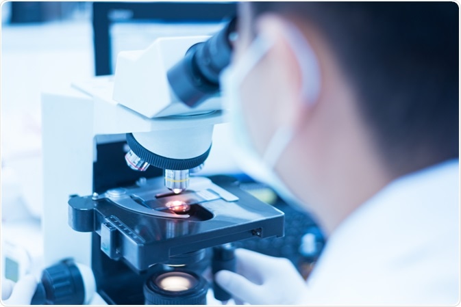
Electron microscopes have long been used to study the ultrastructure of a myriad of biological samples, such as cells, biopsies, microorganisms, large molecules, and more.

Image Credit: Komsan Loonprom/Shutterstock.com
Advances continue to be made in the technology and methodology used in electron microscope techniques which are growing their capabilities.
Recently, a study conducted by scientists at Helmholtz Zentrum Muenchen established a new electron microscopy technique that uses differently sized nanospheres as markers to visualize gene expression of cells.
The need for multi-color genetic labeling in electron microscopy
Electron microscopy (EM) has emerged as the gold standard in terms of methods used to determine cellular processes at the mechanical level, to reveal the architecture of cell circuits. However, in comparison with the wide variety of fluorescent proteins and optical reporters that are available for enhancing other imaging techniques, equivalent genetic tools for use in EM have been limited.
While some semi genetic markers have been developed, the establishment of a fully genetic EM marker proved more difficult to achieve. In 2019, scientists published a paper in the journal ACS nano which describes how a team based in Germany looked for iron-sequestering protein complexes that were of a large enough size to identifiable from the molecules existent within the cell.
Prof. Dr. Gil Gregor Westmeyer, who worked with the team at Helmholtz Zentrum Muenchen, explains that while imaging methods that use fluorescent proteins have proven invaluable to advancing our knowledge of the small structures within cells, their use is limited due to its drawbacks relating to the generally low resolution of light microscopes.
To overcome this resolution, electron microscopes have been employed by some research projects because of their greater resolution, however, Westmeyer noted that there has been a lack of multi-color genetic labeling of cells to use alongside this technology, which has the effect of limiting EM’s ability to distinguish one cell from another.
With this in mind, Westmeyer led the team of scientists at the Institute for Biological and Medical Imaging (IBMI) of Helmholtz Zentrum München in their research to establish an EM equivalent of the multicolored gene reporters that are used in light microscopy.
The team recognized that while imaging at the resolution of the nanometer is becoming increasingly important for biomedical research, particularly for the engineering of molecular machinery and mapping of cellular circuits, there was a distant lack of "multicolor" markers to enhance the EM method.
Replacing color with geometry
Prof. Dr. Gil Gregor Westmeyer along with his colleagues at Helmholtz Zentrum Muenchen had been working for several years with encapsulins, which are tiny, non-toxic proteins extracted from bacteria.
These proteins will automatically assemble nano compartments within a cell, enabling chemical reactions to take place intracellularly without impacting on the metabolism of the cell.
Scientists have demonstrated that they can control the diameters of the nano compartments that are constructed within the cells by altering the experimental conditions via genetic programming. Instead of using colors, as is used by fluorescence microscopy, the team developed a method that instead uses geometry, where different sizes replace different shades.
Therefore, the labeling is dependent on the size of the nanospheres.
To enable a strong contrast to be produced in the images generated by EM, the team used ferroxidase, an enzyme that can become encapsulated within encapsulins.
In the case where the pores of the nano compartments allow iron ions to flow into the interior lumen, divalent iron ions then become oxidized into their trivalent from by ferroxidase. This results in the formation of insoluble iron oxides inside the lumen.
Because metals absorb electrons, in a similar way to which dense bones absorb x-ray radiation, they create good contrasts. This means encapsulins visible in images created with EM.
Investigating how memories are stored
The researchers were able to use their newly established method to visualize and investigate neural circuits. While conventional EM has a very high resolution, it is not able to identify certain neuronal cell types.
However, the team was able to prove that with their new method of EM with differently sized nanospheres as markers, they were able to label specific neuronal cells and create readings of which cell types were making connections as well as reporting on the state of the cells.
While there are a number of applications that may be made possible with the use of this innovation, one particular use that may soon be under development is that of investigating the nature of memories.
Scientists believe that the new technology could be used to reveal how the brain is wired, and potentially uncover how memories become forged in neuronal networks. This may have implications for future treatments of patients suffering from memory loss or other forms of cognitive decline that implicate short term and long term memory.
It could also help us understand why we remember some details and forget others, which could lead to better memorization techniques, which may be useful in some professions.
Sources:
- Beck, M. and Baumeister, W. (2016). Cryo-Electron Tomography: Can it Reveal the Molecular Sociology of Cells in Atomic Detail?. Trends in Cell Biology, 26(11), pp.825-837. https://www.ncbi.nlm.nih.gov/pubmed/27671779
- Sigmund, F., Massner, C., Erdmann, P., Stelzl, A., Rolbieski, H., Desai, M., Bricault, S., Wörner, T., Snijder, J., Geerlof, A., Fuchs, H., Hrabĕ de Angelis, M., Heck, A., Jasanoff, A., Ntziachristos, V., Plitzko, J. and Westmeyer, G. (2018). Bacterial encapsulins as orthogonal compartments for mammalian cell engineering. Nature Communications, 9(1). https://www.nature.com/articles/s41467-018-04227-3#citeas
- Sigmund, F., Pettinger, S., Kube, M., Schneider, F., Schifferer, M., Schneider, S., Efremova, M., Pujol-Martí, J., Aichler, M., Walch, A., Misgeld, T., Dietz, H. and Westmeyer, G. (2019). Iron-Sequestering Nanocompartments as Multiplexed Electron Microscopy Gene Reporters. ACS Nano, 13(7), pp.8114-8123. https://pubs.acs.org/doi/10.1021/acsnano.9b03140
Further Reading