Rafael Fernández-Leiro from the Spanish National Cancer Research Centre (CNIO) led researchers from the Genomic Integrity and Structural Biology Group to discover how specific proteins ensure the rectification of errors introduced into the DNA when it replicates.

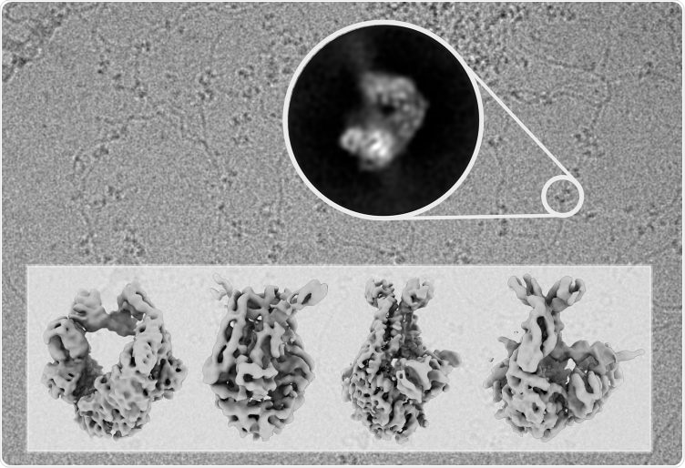
The background of the illustration is a photograph taken with the electron microscope showing DNA molecules decorated with MutS molecules, scanning the DNA for errors. The lower part shows the MutS structures at different stages of the repair process resolved in this study. Image Credit: CNIO.
The researchers used cryo-electron microscopy to render the MutS protein, also called the guardian of the human genome, visible. This allowed them to elucidate how this single protein coordinates the vital DNA repair process from the start to the end.
The research was performed in collaboration with Meindert Lamers of the Leiden University Medical Center (LUMC, The Netherlands) and Titia Sixma of the Netherlands Cancer Institute and the Oncode Institute. The findings have been reported in a paper published in Nature Structural & Molecular Biology.
DNA replication is one of the stages of cell division. During this stage, DNA polymerase reproduces the cell’s genetic information so that it can be transferred to the daughter cell. This is a highly accurate process, but errors can occur sometimes. These errors must be repaired because they can otherwise result in the development of tumors.
Already, the researchers had explained in earlier publications that DNA polymerase features its own repairer, an exonuclease. This enables it to rectify the errors introduced during DNA replication. However, when this corrector is not adequate, the MutS protein plays a role by scanning the copied DNA for errors and then initiating and finishing the repair of any errors it detects.
However, to date, it was not apparent how a single protein coordinates so many different processes. Now, the published international study has successfully uncovered the mechanism.
Using cryo-electron microscopy, we were able to observe MutS while it carried out its functions and capture its molecular structure in successive conformations. With this information, we were able to understand how a single protein can coordinate the whole process, which has to be extremely accurate.”
Rafael Fernández-Leiro, Spanish National Cancer Research Centre
Deep insights into the DNA repair process, which involves the exonuclease, DNA polymerase, and the MutS protein, is crucial to perceive how modifications in any of these proteins result in mutations and, thus, to an elevated level of risk in developing specific types of tumor, like endometrial cancer and Lynch syndrome.
The team highlights that revealing protein structures is possible only because of the vast technological advances in electron microscopy in the recent past.
Electron microscopy allows us to obtain very high-resolution images of proteins as they carry out their functions. With these images, we can reconstruct the three-dimensional structure of the protein in the computer and generate an atomic model that allows us to understand how it works.”
Rafael Fernández-Leiro, Spanish National Cancer Research Centre
Source:
Journal reference:
Fernandez-Leiro, R., et al. (2021) The selection process of licensing a DNA mismatch for repair. Nature Structural & Molecular Biology. doi.org/10.1038/s41594-021-00577-7.