In a recent study led by UCL researchers, the mass of human chromosomes, which hold the instructions for life in practically every cell of the human body, was quantified using X-rays for the first time.

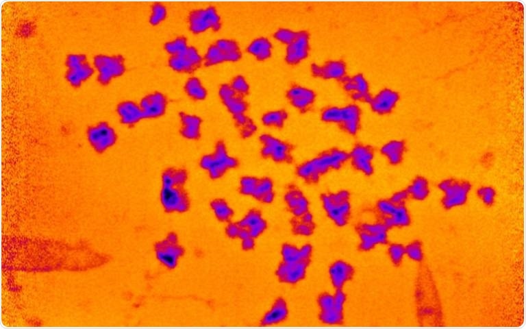
The spread of 46 chromosomes, with artificial color added. Image Credit: Archana Bhatiya et al./Chromosome Research.
Researchers employed a strong X-ray beam at the UK’s national synchrotron facility, Diamond Light Source, to measure the number of electrons in a spread of 46 chromosomes, which they then used to compute mass for the study, which was published in Chromosome Research.
They observed that the chromosomes were nearly 20 times heavier than the DNA they carried—a far bigger amount than previously thought, implying that there may be missing components still to be uncovered.
In addition to DNA, chromosomes contain proteins that perform a range of roles, from reading the DNA to controlling cell division processes, and tightly packing 2-m strands of DNA into our cells.
Chromosomes have been investigated by scientists for 130 years but there are still parts of these complex structures that are poorly understood. The mass of DNA we know from the Human Genome Project, but this is the first time we have been able to precisely measure the masses of chromosomes that include this DNA.”
Ian Robinson, Study Senior Author and Professor, London Centre for Nanotechnology, University College London
“Our measurement suggests the 46 chromosomes in each of our cells weigh 242 picograms (trillionths of a gram). This is heavier than we would expect, and, if replicated, points to unexplained excess mass in chromosomes,” added Robinson.
The researchers employed a technique known as X-ray ptychography to build a very sensitive 3D reconstruction, which involves sewing together the diffraction patterns that occur when the X-ray beam travels through the chromosomes.
Because the beam launched at Diamond Light Source was billions of times brighter than the Sun, an exquisite resolution was attainable (i.e., there was an exceptionally large number of photons passing through at a given time).
The chromosomes were photographed at metaphase, right before they split into two daughter cells. This is the stage at which packaging proteins wrap the DNA into highly compact, accurate structures.
A better understanding of chromosomes may have important implications for human health. A vast amount of study of chromosomes is undertaken in medical labs to diagnose cancer from patient samples. Any improvements in our abilities to image chromosomes would therefore be highly valuable.”
Archana Bhartiya, Study Lead Author and PhD Student, London Centre for Nanotechnology, University College London
At metaphase, each human cell has 23 pairs of chromosomes, for a total of 46. There are four copies of 3.5 billion base pairs of DNA in each of them.
Source:
Journal reference:
Bhartiya, A., et al. (2021) X-ray Ptychography Imaging of Human Chromosomes After Low-dose Irradiation. Chromosome Research. doi.org/10.1007/s10577-021-09660-7.