Recent research carried out by RIKEN biologists on the development of symmetry between the left and right sides in mice embryos is anticipated to provide a deeper understanding of the causes of disease, birth defects, and genetic syndromes.

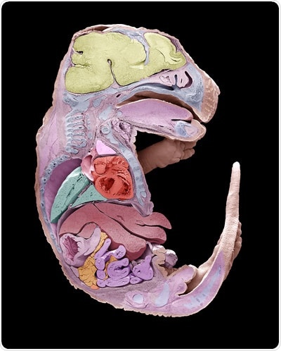
A scanning electron micrograph of one-half of a mouse embryo. The other half would be different in some respects. RIKEN biologists have discovered the role that genes play in the development of this left–right asymmetry. Image Credit: © Steve Gschmeissner/Science Photo Library.
Embryos begin from symmetric bundles of cells and, later on, develop into animals whose right sides vary from their left sides. Developmental biologists intend to find the origins of this left-right asymmetry as such knowledge might provide insights into the basic biology of development, and also the causes of birth defects and genetic syndromes.
The left-right breaking event is the initial step in establishing left-right asymmetry in an embryo. In fish, frog, and mouse embryos, this starts with hair-like cilia producing fluid flow that runs leftward. This fluid flow later downregulates Dand5 mRNA on the left-hand side of the embryo.
Recently, Hiroshi Hamada of the RIKEN Center for Developmental Biology and his group along with scientists from Switzerland and elsewhere in Japan analyzed the factors that suppress Dand5 mRNA.
The researchers initially tracked down the crucial portion of the mRNA by comparing sequences between mammalian genes to unravel the conserved portion. This showed a conserved 200-nucleotide region of the proximal 3′-UTR.
Embryos missing a functional copy of the Bicc1 gene showed defects in left-right patterning, associating the Bicc1 protein with left-right asymmetry. Upon deleting the first exon of Bicc1 with CRISPR–Cas9 gene editing, the resultant mouse embryos were formed symmetrically.
The researchers identified that the Bicc1 protein attaches to the GACGUGAC sequence in the untranslated region of Dand5 mRNA. Additional research revealed that Bicc1 needs to interact with the Cnot3 component of the Ccr4–Not deadenylase complex.
The observations substantially enhance the understanding of the development of left-right asymmetry.
Our study has revealed how cells respond to fluid flow generated by cilia. We have confirmed that left–right symmetry is broken by degradation of a particular mRNA on the left side in response to the directional fluid flow. Discovering the involvement of the RNA degrading system in sensing the fluid flow is an exciting breakthrough.”
Hiroshi Hamada, Center for Developmental Biology, RIKEN
As Bicc1 is involved in kidney disease, Hamada anticipates that a similar process might operate when cells sense other kinds of fluid flows, like kidney epithelial cells identifying urine flow.
As a further step, the researchers intend to examine how calcium activates the Bicc1–Ccr4 complex, particularly if calcium impacts complex formation or phosphorylation of Bicc1 and Ccr4 components.
Source:
Journal reference:
Minegishi, K., et al. (2021) Fluid flow-induced left-right asymmetric decay of Dand5 mRNA in the mouse embryo requires a Bicc1-Ccr4 RNA degradation complex. Nature Communications. doi.org/10.1038/s41467-021-24295-2.