The ImageXpress® Confocal HT.ai High-Content Imaging System makes use of a seven-channel laser light source with eight imaging channels to allow for highly multiplexed assays while maintaining high throughput by using shortened exposure times.
Water immersion objectives help to enhance image resolution and reduce aberrations to ensure scientists can see deeper into thick samples.
The powerful combination of MetaXpress® software and IN Carta® software helps to streamline workflows for advanced phenotypic classification and 3D image analysis with machine learning capabilities and an intuitive user interface.
Introduction to the Organoid Innovation Center and ImageXpress Confocal HT.ai system. Video Credits: Molecular Devices UK Ltd
Advantages
Enable Greater Assay Flexibility
Eight imaging channels with laser excitation allow for greater assay flexibility, higher image brightness, and flexibility to use targeted imaging such as QuickID. Automated Water Immersion objectives provide greater numerical aperture and matched refractive index between the sample and the immersion media for decreased aberrations and improved resolution.
Increase Throughput With Higher Quality Images
Higher excitation power offers shorter exposure times, increased signal, and faster acquisition of 3D samples. The micro-lens enhanced spinning disk confocal gives users a flat field of view for more reproducible and precise image analysis. Shorter exposure times generate up to a two-fold boost in scan speed. FRET experiments that have been carried out using lasers for CFP and YFP have helped to expand research.
Accelerate Analysis Speeds
IN Carta Image Analysis Software carries out complicated segmentation and classification. Phenoglyphs offer a challenging trainable classification, and SINAP offers trainable segmentation for all images. Using MetaXpress® PowerCore Software, users can accelerate analysis speeds by 40× with multi-threaded, parallel processing.
Experiment duration is decreased from hours to minutes as the 3D analysis that serves as a bottleneck is removed.
Features
High-Intensity Laser Light Source
With shorter exposure times, users can capture images faster. Multiplex experiments using seven lasers and eight imaging channels.
Accurate 3D Measurements
Optimized for confocal imaging, Custom Module Editor 3D analysis allows for 3D distance and volume measurements.
Wide Field of View
A wide field of view eliminates missed targets and allows whole-well confocal imaging. Next-generation dual microlens-enhanced spinning disk technology offers a large, flat field of view for more precise and reproducible analysis.
Exclusive AgileOptix™ Spinning Disk Technology
The Exclusive AgileOptix™ Spinning Disk Technology offers increased sensitivity with dedicatedly designed optics, high-powered laser illumination, and an sCMOS sensor. Swappable disk geometries offer flexibility between resolution and speed.
IN Carta Image Analysis Software
It leverages machine learning to enhance the precision and strength of high-content image analysis, thereby supplying data insights that other technologies cannot. The complexity associated with image analysis is removed, resulting in a modern user interface with intuitive guided workflows.
Multiple Imaging Modes
With water immersion optics as a standard option, the system provides phase contrast and brightfield label-free imaging, widefield, and confocal fluorescent imaging.
Automated Water Immersion Objective Technology
Offers greater image resolution and sensitivity with up to 4x increase in signal leading to lower exposure times.
Wide Dynamic Range
Quantifies low and high-intensity signals in a single image with >3 log dynamic range intensity detection.
AgileOptix™ Technology
AgileOptix technology combines a powerful solid-state light engine, a scientific CMOS sensor, custom optics, and the ability to change between five different disk geometries.
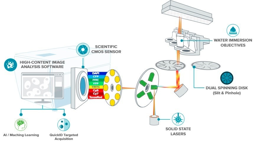
Image Credit: Molecular Devices UK Ltd
Images Taken With Same Exposure Times Show Different Average Intensities
High-output laser excitation can minimize exposure time by a maximum of 75%. The 8-channel laser light source outputs 1,000 mW/channel and contains near IR — which is ideal for users with increased multiplexing needs.
- Produce up to a two-fold boost in scan speed attributed to considerably minimized exposure times.
- Achieve sharper images with higher signal-to-noise.
- Run FRET experiments with lasers for YFP and CFP with increased multiplexing requirements.

Cy5. Image Credit: Molecular Devices UK Ltd
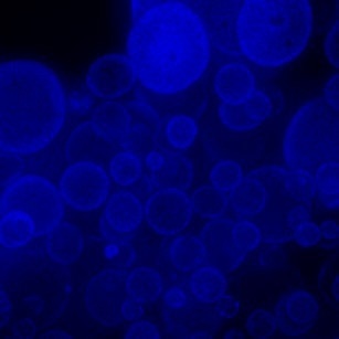
DAPI. Image Credits Molecular Devices UK Ltd
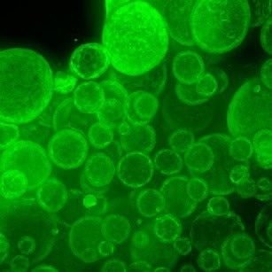
FITC. Image Credit: Molecular Devices UK Ltd
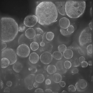
TRITC. Image Credit: Molecular Devices UK Ltd
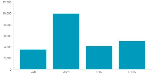
Average intensity for same exposure time: Laser. Image Credit: Molecular Devices UK Ltd
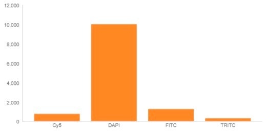
Average intensity for same exposure time: LED. Image Credit: Molecular Devices UK Ltd
IN Carta Image Analysis Software
Efficient analytics integrated with an intuitive user interface ease workflows for phenotypic profiling and image analysis. Cutting-edge features offer the functionality users require to explore data in 2D, 3D, and 4D — at scale — and supply real-time insights without the need for complicated pre- or post-processing operations.
Enhance the specificity of image analysis workflows with the SINAP deep-learning module and see that Segmentation is not an issue. Using machine learning, conduct complex phenotypic analysis within a user-friendly Phenoglyphs module.
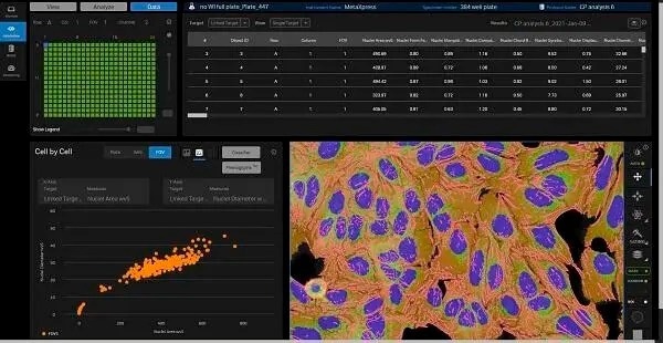
Image Credit: Molecular Devices UK Ltd
Cellular Image Gallery
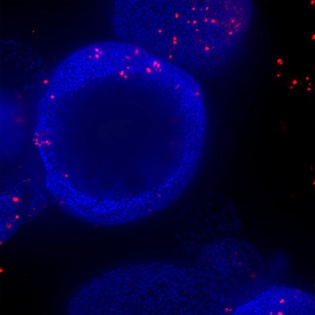
HT.ai Organoid with Black Hole. Image Credit: Molecular Devices UK Ltd.
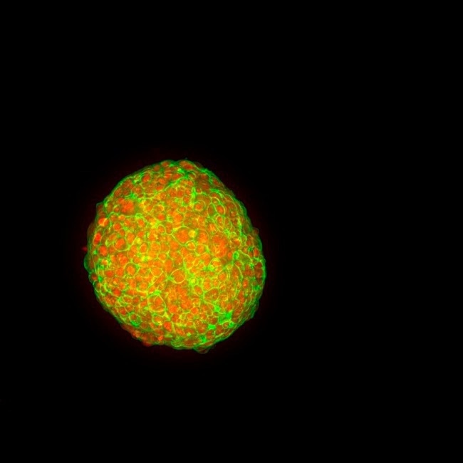
HT.ai Organoid overlay z-steps. Image Credit: Molecular Devices UK Ltd.
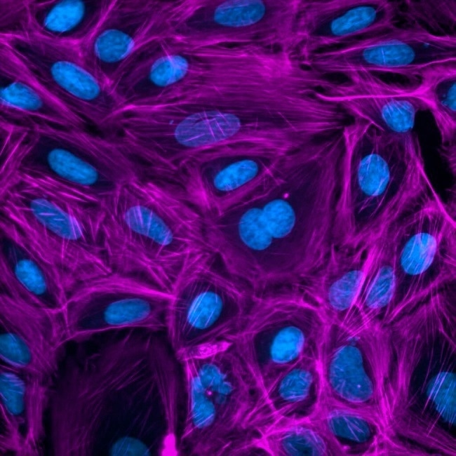
HT.ai Cell Painting. Image Credit: Molecular Devices UK Ltd.
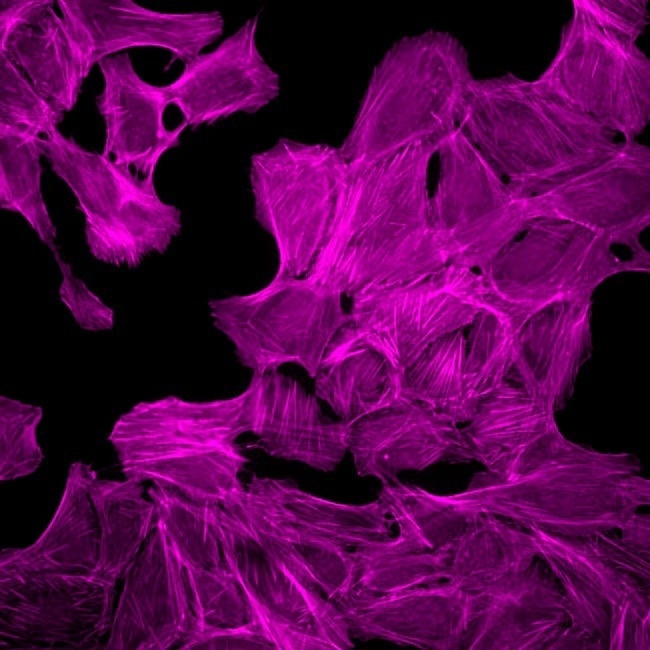
HT.ai Organoid-3. Image Credit: Molecular Devices UK Ltd.
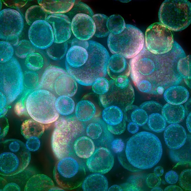
HT ai organoid encoded. Image Credit: Molecular Devices UK Ltd.
†Data and images were acquired during development using customer samples. Results may vary. Highlighted features’ price, time to deliver, and specifications will vary based on mutually agreed technical requirements. Solution requirements may cause adjustment to standard performance.