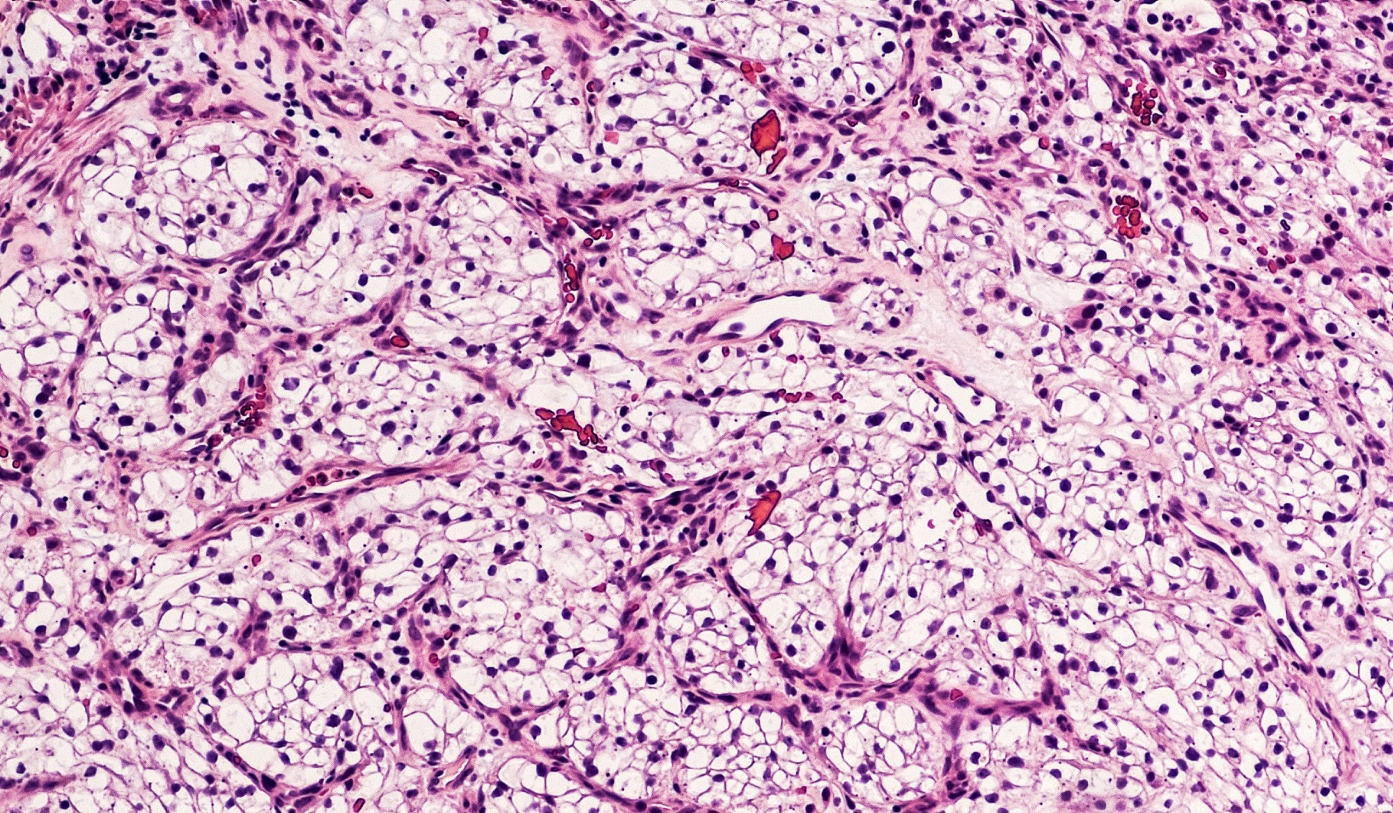
Over 80% of renal cancer cases are clear cell renal cell carcinoma or ccRCC, which shows no obvious symptoms in the early stages and for which, currently, no early screening serum biomarkers exist.
In a recent study published in Heliyon, scientists used transcriptomics and radiomics data clustering and its association with molecular characteristics, clinical pathologies, and prognosis to define intrinsic subtypes of ccRCC.
 Study: Identification of clear cell renal cell carcinoma subtypes by integrating radiomics and transcriptomics. Image Credit: David A Litman/Shutterstock.com
Background
Although ccRCC is the most prevalent of all malignant tumors of the kidneys, the early symptoms of ccRCC are minimal and difficult to discern.
The lack of any serum biomarkers to screen for ccRCC also results in the detection of cancer only in the advanced stages in over 25% of the patients, after which a combination of targeted therapy, immunotherapy, and surgery are the only recommended treatment options.
Current methods of predicting treatment outcomes include a percutaneous biopsy, which presents the risk of needle tract metastasis, bleeding, and infections.
Furthermore, the individual prognosis for ccRCC patients is substantially heterogeneous, and small tissue biopsies pose the danger of not accurately representing this heterogeneity.
Moreover, conventional detection methods such as magnetic resonance imaging, computed tomography, and ultrasound cannot distinguish between varying pathologies quantitatively.
About The Study
In the present study, the researchers used radiotranscriptomics and unsupervised clustering to evaluate molecular, immune, prognostic, and clinicopathological characteristics of ccRCC and identify the various intrinsic subtypes.
The field of radio transcriptomics combines the quantitative image analysis features of radiomics with the detailed molecular information and insights into clinical pathology gained through transcriptomics while overcoming the shortcomings of each field.
Through the integration of genomic or transcriptomic data and radiomics, the method provides a non-invasive but efficient method to predict the tumor microenvironment.
Previous studies, including some by the same team of researchers, had used radiotranscriptomic analyses to determine metabolism, proliferation, and immune activation-based subtypes of non-small-cell lung cancer.
Here, the researchers also conducted a multi-omics-based comprehensive investigation of the functional aspects of the genes identified to have clinical significance in ccRCC.
The present retrospective study consisted of a training and a validation cohort into which patients were enrolled if they had been diagnosed with ccRCC, had solitary lesions, and had undergone contrast-enhanced computed tomography or CECT imaging.
Information on the clinicopathological parameters, as well as transcriptomic data, were obtained for both cohorts.
Regions of interest were manually identified from the 3D CECT images, and areas of hemorrhage, necrosis, calcifications, and cystic changes representing internal tumor radiomics features were delineated.
107 radiomics features, including first-order, shape, gray-level dependence matrix, and four more types of features, were identified.
The transcriptomic data and the radiomic features were standardized and scaled during preprocessing, and 300 genes that were found to be highly variable were identified from the transcriptomic data for the clustering analysis. Tumor subtypes were then identified using unsupervised clustering analyses.
Subsequently, immune, molecular, prognostic, and clinicopathological characteristics of the various tumor subtypes were examined through comparisons of disease-free survival, overall survival, tumor grade, and stage, as well as the age and sex of the patients.
Molecular and transcriptomic features such as signaling pathways, differential gene expression, immune function checkpoints, and immune cell infiltration were also compared across the subtypes.
Additionally, comparisons of gene mutation profiles, microsatellite instability, tumor mutational burden, tumor immune dysfunction, and ribonucleic acid (RNA) interference were conducted. The radiomics data from the validation cohort was then used to validate the different tumor subtypes.
Major Findings
The study identified three ccRCC subtypes, namely C1, C2, and C3, with substantial distinctions in immune, molecular, prognostic, and clinicopathological characteristics. Subtype C2 showed better survival outcomes than C1 and C3, differing significantly in the prognostic characteristics.
Analyses of immune characteristic differences showed that the C2 subtype had significant metabolic pathway activation. In contrast, the C1 subtype had higher infiltrating immune cells and function and elevated levels of immune checkpoint molecules.
Gene mutation analyses identified the Von Hippel-Lindau tumor suppressor (VHL) and the Polybromo-1 (PBRM1) genes as the two most common genes that were mutated in ccRCC. The C3 subtype had more mutated genes than the other two subtypes.
Although higher levels of immune dysfunction in tumors were observed in subtypes C1 and C3, all the subtypes showed similar microsatellite instability, RNA interference, and tumor mutation burden. Furthermore, the data from the validation cohort corroborated the prognostic and clinicopathological features for the three subtypes.
Conclusions
To summarize, the study used a radiotranscriptomics approach with unsupervised clustering to define three intrinsic ccRCC subtypes that could be used to characterize the prognostic and clinicopathological features of ccRCC and used for non-invasive risk stratification.
These subtypes provide an efficient method to evaluate the heterogeneity of ccRCC, which can be used for effective and personalized cancer treatment.
Journal reference:
-
Gao, R., Pang, J., Lin, P., Wen, R., Wen, D., Liang, Y., Ma, Z., Liang, L., He, Y., & Yang, H. (2024). Identification of clear cell renal cell carcinoma subtypes by integrating radiomics and transcriptomics. Heliyon, 10(11), e31816. doi:https://doi.org/10.1016/j.heliyon.2024.e31816. https://www.sciencedirect.com/science/article/pii/S2405844024078472