Overview
Cryopreserved primary mouse hepatocytes are co-cultured with non-parenchymal stromal cells in HUREL® Mouse™.
Example Metabolic Activity (nmoles/hr/106 cells). Source: Visikol Inc.
| Substrate |
Enzyme |
Concentration (µM) |
Day 1 |
Day 4 |
Day 8 |
| 7-Ethoxycoumarin |
Phase I |
100 |
0.034 |
0.017 |
0.016 |
| 7-Hydroxycoumarin |
Phase II |
100 |
51.5 |
46.5 |
0.21 |
| 7-Hydroxycoumarin |
Phase III |
100 |
1.76 |
1.02 |
0.08 |
Primary mouse hepatocytes that had been frozen were thawed, plated on HUREL PlatinumHeps™ Media supplemented with 10% serum, and then switched to HUREL PlatinumHeps™ Basal Media 24 hours later.
The concentrations of CYP substrates are listed in the table above, and metabolite formation is expressed as nmoles/hr/106 cells. On Days D1, D4, and D8, after cell delivery, all incubations were performed in triplicate and incubated for 60 minutes.
The reactions happened in a humid incubator with 5% CO2 at 37 °C. Supernatants collected were kept at –20 °C until an additional LC/MS/MS analysis.

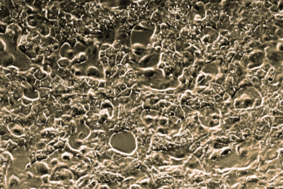
Phase contrast image in a 24-well at a 10× magnification. Image Credit: Visikol Inc.

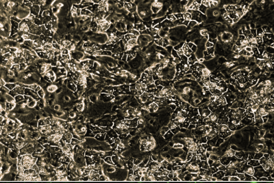
Phase contrast image in a 24-well at a 10× magnification. Image Credit: Visikol Inc.

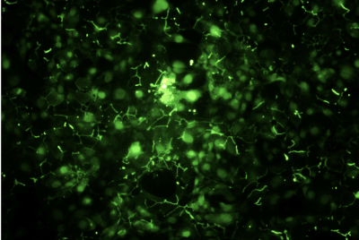
Bile canaliculi assayed via 5-(and-6)-carboxy-2’, 7’-dichlorofluorescein diacetate (C-DCFDA) stain at a concentration of 5 µM and imaged in the GFP channel in a 96-well at 10x magnification with filters EX/EM 492-495/512-527 nm. Image Credit: Visikol Inc.
Example culture origin
Animal donor demographics
- Strain: CD-1
- Number of Donors: 5
- Age (years)
- Sex: Male