Overview
MatTek’s newly advanced 3D full-thickness model of human skin takes preclinical testing to the next level by providing a physiological means to assess early molecular markers of potency.
The growth of innovative skincare products needs improved, human-relevant in vitro testing technologies. While world-leading personal care and cosmetic scientists assess novel ingredients and formulations, they select the EpiDermFT 3D human skin model.
Key features
- Big donor inventory with matched Keratinocytes and Fibroblasts
- Delivered user-friendliness
- Comes with adult or neonatal donors
Technology
Engineered to enable in vitro skin research in which fibroblast-keratinocyte cell interactions are significant, MatTek’s EpiDermFT system comprises normal, human epidermal keratinocytes (NHEK) and normal, human dermal fibroblasts (NHFB) cultured to develop a multilayered model of the human epidermis and dermis.
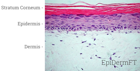
Image Credit: MatTek
Having been cultured at the air-liquid interface in simple-to-handle tissue culture inserts, EpiDermFT achieves levels of differentiation at the cutting edge of tissue engineering technology.
EpiDermFT comprises organized involucrin, keratin 5 expressing basal cells, keratin 10 expressing spinous and granular layers, and cornified epidermal layers analogous to those found In vivo.
The dermal compartment is made of a collagen matrix consisting of viable normal human dermal fibroblasts (NHDF). The epidermal and dermal layers are mitotically and metabolically active and display in vivo-like morphological and growth characteristics, which are even and highly reproducible.
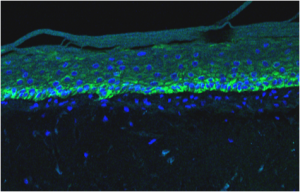
Keratin 5. Image Credit: MatTek
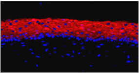
Keratin 10. Image Credit: MatTek
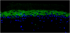
Involucrin. Image Credit: MatTek
A well-developed basement membrane is present at the dermal or epidermal junction. Lamina lucida, hemidesomosomes (›), lamina densa (→), and anchoring fibril structures (») are obvious by transmission electron microscopy.

Image Credit: MatTek
Human relevance
A robust skin model with various applications for assessing dermal care product efficiency and toxicity. Formulations are implemented to the apical tissue surface, just like in vivo.
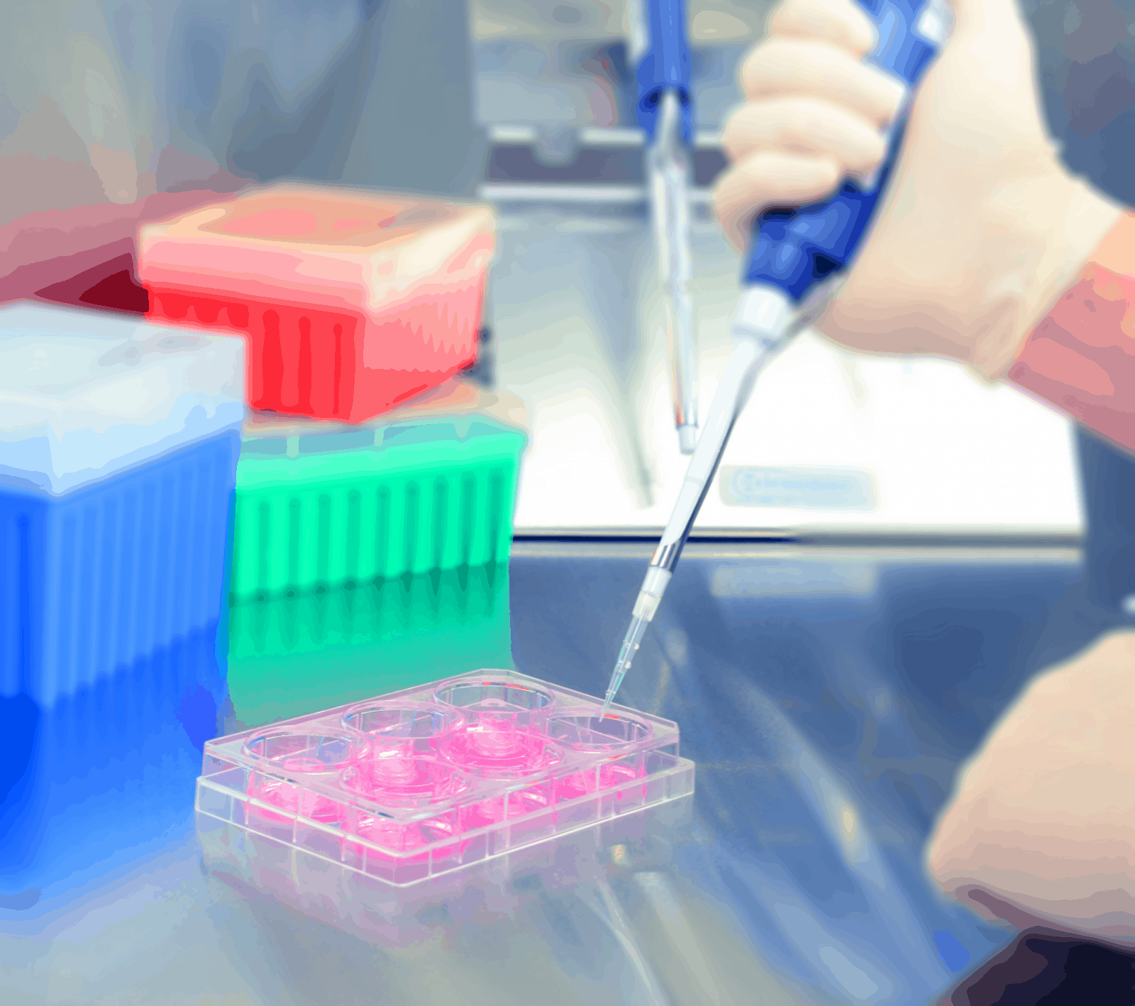
Image Credit: MatTek
Applications
EpiDermFT tissues are utilized throughout an extensive range of applications such as scientific efficacy and claims substantiation. As a result of a dry skin surface, EpiDermFT could be utilized to assess biological responses to topical applications of formulations or ingredients. Easy protocols and the assessment of early molecular endpoints enable research to obtain data in days, not weeks, or months.
Anti-aging
Analyze variations in extracellular matrix gene and protein expression and histological structure to identify the anti-aging efficacy of cosmetic ingredients or eventual formulation. It comes from EpiDermFT from a range of adult donors.
Wound healing
Users can utilize EpiDermFT to assess therapeutics with the wound healing assay kit. Wound healing assay kits consists of 24 pre-wounded tissues and comes with epidermal-only wounds or full-thickness wounds of the dermis and epidermis.
Skin hydration
Utilize EpiDermFT to evaluate the efficacy of topically applied moisturizers by quantifying the epidermis’ electrical impedance. Amenable to gel, creams, lotions, or other formulations, the skin hydration assay enables data analysis within 24 hours.
UV protection
Users can analyze cyclopyrimidine dimer formation, inflammatory mediator release, and oxidative endpoints to assess the UV protective efficacy of formulations or compounds.