Overview
The advanced 3D in vitro co-culture skin model from MatTek tends to integrate human melanocytes into a highly-differentiated epidermal tissue. Assess novel skin-lightening agents in vitro with the foremost pigmented skin model.
MelanoDerm is being used by cosmetic scientists all over the world to evaluate skin brightening agents and formulations. Minimize clinical testing time and costs by screening compounds and formulations in MatTek’s MelanoDerm 3D tissue model.
Features
- Distinguished 3D co-culture of melanocytes and keratinocytes
- Melanocytes from different ethnicities are available
- Quantitative readouts can be done within 2 weeks
- Delivered user-friendly
Technology
MatTek’s MelanoDerm System consists of normal, human-derived epidermal keratinocytes (NHEK) and melanocytes (NHM) cultured to develop a multilayered, highly distinguished model of the human epidermis.
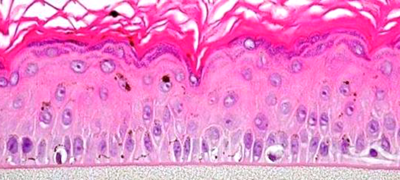
Image Credit: MatTek
The NHM within co-cultures experiences melanogenesis as a result of tissue pigmentation. MelanoDerm has been produced with the help of a serum-free medium in the absence of artificial stimulators of melanogenesis like IBMX and TPA.
Image Credit: MatTek
The cultures have been grown on tissue culture inserts at the air-liquid interface, enabling for topical application of self-tanning agents or skin lighteners. MelanoDerm offers a beneficial in vitro means to assess cosmetic and pharmaceutical agents that have been developed to modulate skin pigmentation.
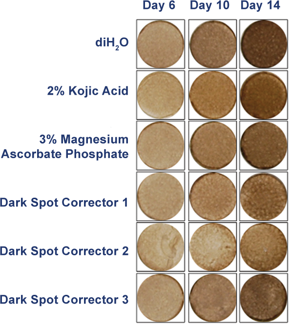
Image Credit: MatTek
MelanoDerm displays in vivo-like morphological and ultrastructural characteristics which are even and highly reproducible. In the basal cell layer of MelanoDerm, NHM is dendritic and concurrently produces melanin granules which increasingly populate the tissue layers.
While being cultured for up to three weeks, MelanoDerm tissues turn out to be highly pigmented with retention of normal epithelial morphology. Tissues consisting of NHM derived from black donors display increased pigmentation compared to those consisting of Caucasian-derived or Asian-derived NHM; all three kinds of cultures are evidently darker compared to NHM-free tissues (EpiDermTM).
The topical application of well-known inhibitors of melanogenesis considerably decreases macroscopic darkening and melanin production. On the contrary, NHM present inside the tissue will react to identified stimulants of melanogenesis, like α-melanocyte-stimulating hormone and β-fibroblast growth factor, to produce tissues that have the potential to darken faster compared to controls that are left untreated.
For almost two decades, MelanoDerm’s direct assays have been integrated as a crucial testing strategy in laboratories. Having dozens of technical references ready, this increasing body of data illustrates that MelanoDerm offers an affordable and effective way of evaluating skin pigmentation while preventing species extrapolation and also the use of laboratory animals.
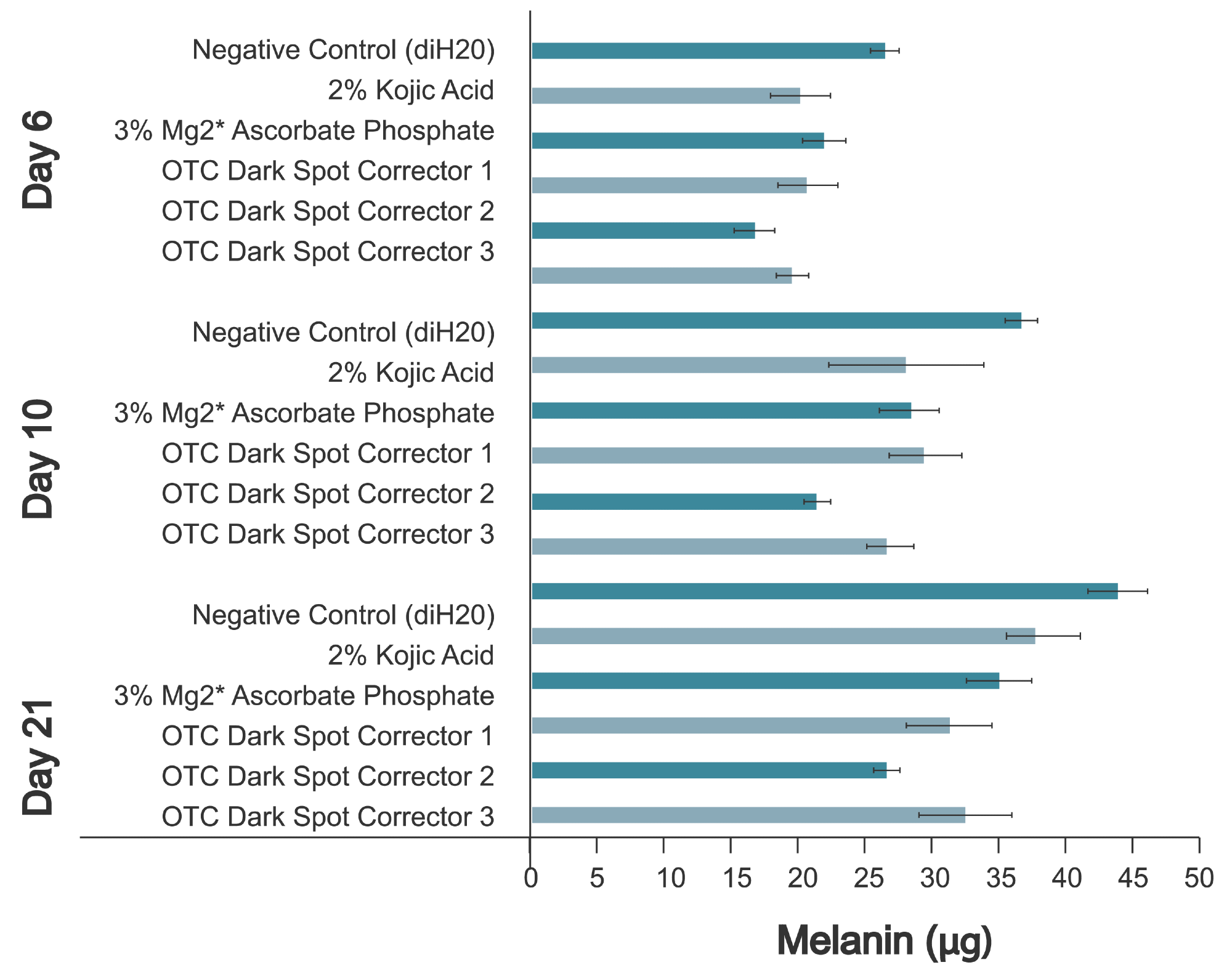
Image Credit: MatTek

Image Credit: MatTek
Human relevance
Assess topical formulations or actives for skin-lightening efficiency with the human cell-based MelanoDerm tissue model.
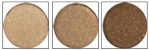
Image Credit: MatTek
Applications
MelanoDerm offers a low-cost and efficient means of evaluating skin pigmentation while preventing species extrapolation and the use of laboratory animals. Easy protocols and the assessment of early cellular endpoints enable researchers to obtain data just in days, not weeks or months.
Skin brightening
A quantitative assay available to identify melanin content in the tissue is executed with the help of the SOLVABLE™ melanin assay. This assay could be utilized to test the skin-brightening efficacy of eventual cosmetic formulations.
Pigmentation studies
MelanoDerm can be employed to investigate the physiological mechanisms of skin pigmentation.