Overview
Pharmaceutical and personal care companies depend on MatTek’s oral mucosal models to assess their raw materials and eventual product formulations.
Technology
Several toxicology and industrial laboratories are actively looking for alternatives for high-cost clinical or whole-animal testing. Personal care, oral care, and pharmaceutical companies have launched in vitro toxicology testing to assess their raw materials and final product formulations.
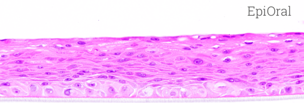
Image Credit: MatTek
MatTek’s EpiOral and EpiGingival tissues comprise normal, human-derived oral epithelial cells. The cells have been cultured to develop multilayered, highly differentiated models of gingival (EpiGingival) and the human buccal (EpiOral) phenotypes.
Especially, the tissues have been cultured on prepared cell culture inserts with the help of serum-free medium and achieve levels of differentiation on the cutting edge of in vitro cell culture technology. The EpiOral and EpiGingival tissue models display in vivo-like morphological and growth characteristics which are even and highly reproducible.
Both EpiGingival and EpiOral express cytokeratin K13 in all layers besides the most apical ones and weakly express cytokeratin K14 in the tissue’s upper layers (Figure 1). Also, the tissues produce naturally occurring antimicrobial peptides known as human beta-defensins (HBDs).
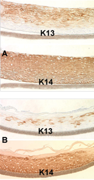
Figure 1. Immuno-staining of: A) EpiOral and B) EpiGingival tissue for cytokeratin K13 and K14. Image Credit: MatTek
EpiOral constitutively expresses HBD-1 and HBD-3 but not HBD-2 (Figure 2).
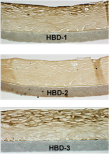
Figure 2. Immuno-staining of the ORL-200 tissue for human beta defensins (HBDs). Image Credit: MatTek
EpiGingival faintly expresses HBD-3 in all layers excluding the uppermost ones and HBD-1 in the apical layers but fails to express HBD-2 (Figure 3).
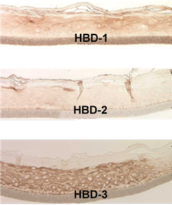
Figure 3. Immuno-staining of the GIN-100 tissue for human beta defensins (HBDs). Image Credit: MatTek
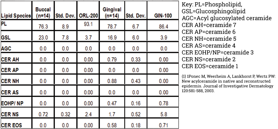
Lipid analysis of the explant tissues together with the EpiGingival (GIN-100) and EpiOral (ORL-200) is displayed below in Table 1. EpiOral and buccal explants consist only Ceramide 2 (Ceramide NS). EpiGingival and gingival tissue comprise Ceramides 1, 2, and 3 (also spoken of as Ceramides NS, EOS, and EOHP/NP, in a respective manner.
Table 1. Lipid analysis of in vivo and in vitro tissues. The in vivo buccal and gingival values are averages for 14 tissue samples. The in vitro tissue values are from a single lot. Source: MatTek
An increasing body of data denotes that EpiOral and EpiGingival efficiently offer an affordable, non-animal means to evaluate toxicology, oral irritation, and pathology-related issues. A representative dose-response curve for the effect of sodium dodecyl sulfate (SDS), a common surfactant utilized in toothpaste, has been displayed in Figure 4.
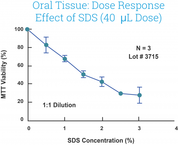
Figure 4. Effect of SDS solutions (40L) on EpiOral (ORL-200) tissue viability following exposure for 1 hour. SDS concentrations were chosen to be in range normally present in toothpaste (0.0 – 3.0%). Image Credit: MatTek
Human relevance
MatTek’s oral tissue models offer highly reproducible, physiological ways to screen recently made oral care products and are beneficial to examine the drug delivery, innate immunity, and pathology of the oral mucosa.
Image Credit: MatTek
Applications
Mucosal and gingival irritation
Quntify gingival or oral epithelial tissue viability and cytokine release with the use of a commercially available ELISA kit or simple MTT assay, and gain ultimate outcomes in a few days.
Drug delivery
EpiOral and EpiGingival are beneficial in finding the absorption potential of the ultimate formulations in vitro.
Model for oral pathologies
EpiOral has been employed in examining oral mucositis, oral candidiasis, and the anti-microbial features of the oral mucosa.